Yuenan Wang, Fada Guan, Siyuan Wang, Wanwei Jian, Mohammad Khan
{"title":"纵向计算机断层扫描(CT)在图像引导小鼠精确放疗中的放射性肝损伤(RILI)评价。","authors":"Yuenan Wang, Fada Guan, Siyuan Wang, Wanwei Jian, Mohammad Khan","doi":"10.1002/pro6.1244","DOIUrl":null,"url":null,"abstract":"<p><strong>Purpose: </strong>We aim to perform image-guided, dose-escalated, well-controlled liver-irradiated animal studies and subsequently evaluate radiation induced liver injury (RILI) using longitudinal CT.</p><p><strong>Methods: </strong>Eighteen 6-8 weeks mice were divided into three groups: control, 15Gy and 30Gy irradiated groups. The animal protocol was approved by the animal care ethics committee of our institution. Precision radiotherapy started with CT simulation, followed by treatment planning using volumetric modulated arc therapy (VMAT), image guidance with cone beam CT (CBCT) and radiation delivery on a medical linear accelerator. Weekly CT was conducted on the same CT simulator using same scanning parameters. At the end of fifth week, all mice were sacrificed, and histological staining was performed. Body weight, liver volume, HU values and histogram distributions were analyzed.</p><p><strong>Results: </strong>Body weight of irradiation groups was significantly reduced compared to that of the control group (<i>p</i><0.05). Liver volume in irradiated groups was reduced too. The average liver HU was significantly reduced in irradiated groups (HU mean = 62±3, 48±6, and 36±8 for the control, 15Gy and 30Gy respectively; <i>p</i> <sub>control vs. 15Gy</sub> < 0.05, <i>p</i> control vs. 30Gy < 0.05). A linear relationship between liver HU and radiation dose was found. Furthermore, HU histogram changes with time and dose showed not only density but also structure might be affected by radiation. HE and Masson Trichrome staining confirmed histological change and increased collagen deposition in irradiated liver.</p><p><strong>Conclusion: </strong>Longitudinal unenhanced CT is a useful imaging tool to evaluate the severity and progression of radiation induced liver injury.</p>","PeriodicalId":32406,"journal":{"name":"Precision Radiation Oncology","volume":"8 4","pages":"182-190"},"PeriodicalIF":2.1000,"publicationDate":"2024-11-09","publicationTypes":"Journal Article","fieldsOfStudy":null,"isOpenAccess":false,"openAccessPdf":"https://www.ncbi.nlm.nih.gov/pmc/articles/PMC11934901/pdf/","citationCount":"0","resultStr":"{\"title\":\"Radiation induced liver injury (RILI) evaluation using longitudinal computed tomography (CT) in image-guided precision murine radiotherapy.\",\"authors\":\"Yuenan Wang, Fada Guan, Siyuan Wang, Wanwei Jian, Mohammad Khan\",\"doi\":\"10.1002/pro6.1244\",\"DOIUrl\":null,\"url\":null,\"abstract\":\"<p><strong>Purpose: </strong>We aim to perform image-guided, dose-escalated, well-controlled liver-irradiated animal studies and subsequently evaluate radiation induced liver injury (RILI) using longitudinal CT.</p><p><strong>Methods: </strong>Eighteen 6-8 weeks mice were divided into three groups: control, 15Gy and 30Gy irradiated groups. The animal protocol was approved by the animal care ethics committee of our institution. Precision radiotherapy started with CT simulation, followed by treatment planning using volumetric modulated arc therapy (VMAT), image guidance with cone beam CT (CBCT) and radiation delivery on a medical linear accelerator. Weekly CT was conducted on the same CT simulator using same scanning parameters. At the end of fifth week, all mice were sacrificed, and histological staining was performed. Body weight, liver volume, HU values and histogram distributions were analyzed.</p><p><strong>Results: </strong>Body weight of irradiation groups was significantly reduced compared to that of the control group (<i>p</i><0.05). Liver volume in irradiated groups was reduced too. The average liver HU was significantly reduced in irradiated groups (HU mean = 62±3, 48±6, and 36±8 for the control, 15Gy and 30Gy respectively; <i>p</i> <sub>control vs. 15Gy</sub> < 0.05, <i>p</i> control vs. 30Gy < 0.05). A linear relationship between liver HU and radiation dose was found. Furthermore, HU histogram changes with time and dose showed not only density but also structure might be affected by radiation. HE and Masson Trichrome staining confirmed histological change and increased collagen deposition in irradiated liver.</p><p><strong>Conclusion: </strong>Longitudinal unenhanced CT is a useful imaging tool to evaluate the severity and progression of radiation induced liver injury.</p>\",\"PeriodicalId\":32406,\"journal\":{\"name\":\"Precision Radiation Oncology\",\"volume\":\"8 4\",\"pages\":\"182-190\"},\"PeriodicalIF\":2.1000,\"publicationDate\":\"2024-11-09\",\"publicationTypes\":\"Journal Article\",\"fieldsOfStudy\":null,\"isOpenAccess\":false,\"openAccessPdf\":\"https://www.ncbi.nlm.nih.gov/pmc/articles/PMC11934901/pdf/\",\"citationCount\":\"0\",\"resultStr\":null,\"platform\":\"Semanticscholar\",\"paperid\":null,\"PeriodicalName\":\"Precision Radiation Oncology\",\"FirstCategoryId\":\"1085\",\"ListUrlMain\":\"https://doi.org/10.1002/pro6.1244\",\"RegionNum\":0,\"RegionCategory\":null,\"ArticlePicture\":[],\"TitleCN\":null,\"AbstractTextCN\":null,\"PMCID\":null,\"EPubDate\":\"2024/12/1 0:00:00\",\"PubModel\":\"eCollection\",\"JCR\":\"Q4\",\"JCRName\":\"Medicine\",\"Score\":null,\"Total\":0}","platform":"Semanticscholar","paperid":null,"PeriodicalName":"Precision Radiation Oncology","FirstCategoryId":"1085","ListUrlMain":"https://doi.org/10.1002/pro6.1244","RegionNum":0,"RegionCategory":null,"ArticlePicture":[],"TitleCN":null,"AbstractTextCN":null,"PMCID":null,"EPubDate":"2024/12/1 0:00:00","PubModel":"eCollection","JCR":"Q4","JCRName":"Medicine","Score":null,"Total":0}
Radiation induced liver injury (RILI) evaluation using longitudinal computed tomography (CT) in image-guided precision murine radiotherapy.
Purpose: We aim to perform image-guided, dose-escalated, well-controlled liver-irradiated animal studies and subsequently evaluate radiation induced liver injury (RILI) using longitudinal CT.
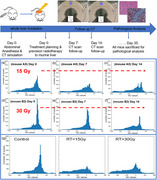
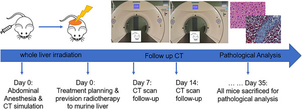
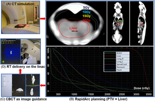
Methods: Eighteen 6-8 weeks mice were divided into three groups: control, 15Gy and 30Gy irradiated groups. The animal protocol was approved by the animal care ethics committee of our institution. Precision radiotherapy started with CT simulation, followed by treatment planning using volumetric modulated arc therapy (VMAT), image guidance with cone beam CT (CBCT) and radiation delivery on a medical linear accelerator. Weekly CT was conducted on the same CT simulator using same scanning parameters. At the end of fifth week, all mice were sacrificed, and histological staining was performed. Body weight, liver volume, HU values and histogram distributions were analyzed.
Results: Body weight of irradiation groups was significantly reduced compared to that of the control group (p<0.05). Liver volume in irradiated groups was reduced too. The average liver HU was significantly reduced in irradiated groups (HU mean = 62±3, 48±6, and 36±8 for the control, 15Gy and 30Gy respectively; pcontrol vs. 15Gy < 0.05, p control vs. 30Gy < 0.05). A linear relationship between liver HU and radiation dose was found. Furthermore, HU histogram changes with time and dose showed not only density but also structure might be affected by radiation. HE and Masson Trichrome staining confirmed histological change and increased collagen deposition in irradiated liver.
Conclusion: Longitudinal unenhanced CT is a useful imaging tool to evaluate the severity and progression of radiation induced liver injury.

 求助内容:
求助内容: 应助结果提醒方式:


