{"title":"腹部创伤分丸ct与双期ct图像质量的比较。","authors":"Shubham Gautam, Anuradha Sharma, Charu Paruthi, Rohini Gupta Ghasi, Krishna Bhardwaj","doi":"10.5114/pjr/200756","DOIUrl":null,"url":null,"abstract":"<p><strong>Purpose: </strong>To compare the image quality in single-pass split-bolus abdominal computed tomography (CT) and conventional biphasic CT in abdominal trauma patients.</p><p><strong>Material and methods: </strong>Sixty-six consecutive abdominal trauma patients referred for CT were randomised into 2 groups: the study group (<i>n</i> = 33), scanned using the split-bolus technique; and the control group (<i>n</i> = 33), scanned using the conventional biphasic technique. CT image quality was analysed subjectively by 2 observers based on a 5-point Likert scale. The images were also analysed quantitatively for attenuation values achieved by region of interest (ROI) placements in major arteries, veins, and solid organs. In addition, the radiation dose in terms of the dose length product (DLP) was compared between the 2 groups.</p><p><strong>Results: </strong>The image quality in both groups ranged from good to excellent in most cases. There was no statistically significant difference in subjective image quality in both the groups as assessed by Likert score. Attenuation values in solid organs and major venous structures were significantly higher in the split-bolus group (<i>p</i> < 0.001). Arterial attenuation values were significantly higher in the control group (<i>p</i> < 0.001), but diagnostic levels were achieved in all patients. There was a reduction of 31.1% in DLP in the split-bolus group.</p><p><strong>Conclusions: </strong>The split-bolus technique offers comparable image quality and higher solid organ and venous enhancement than conventional biphasic protocol at a reduced radiation dose.</p>","PeriodicalId":94174,"journal":{"name":"Polish journal of radiology","volume":"90 ","pages":"e151-e160"},"PeriodicalIF":0.0000,"publicationDate":"2025-03-31","publicationTypes":"Journal Article","fieldsOfStudy":null,"isOpenAccess":false,"openAccessPdf":"https://www.ncbi.nlm.nih.gov/pmc/articles/PMC12049156/pdf/","citationCount":"0","resultStr":"{\"title\":\"Comparison of image quality of split-bolus computed tomography versus dual-phase computed tomography in abdominal trauma.\",\"authors\":\"Shubham Gautam, Anuradha Sharma, Charu Paruthi, Rohini Gupta Ghasi, Krishna Bhardwaj\",\"doi\":\"10.5114/pjr/200756\",\"DOIUrl\":null,\"url\":null,\"abstract\":\"<p><strong>Purpose: </strong>To compare the image quality in single-pass split-bolus abdominal computed tomography (CT) and conventional biphasic CT in abdominal trauma patients.</p><p><strong>Material and methods: </strong>Sixty-six consecutive abdominal trauma patients referred for CT were randomised into 2 groups: the study group (<i>n</i> = 33), scanned using the split-bolus technique; and the control group (<i>n</i> = 33), scanned using the conventional biphasic technique. CT image quality was analysed subjectively by 2 observers based on a 5-point Likert scale. The images were also analysed quantitatively for attenuation values achieved by region of interest (ROI) placements in major arteries, veins, and solid organs. In addition, the radiation dose in terms of the dose length product (DLP) was compared between the 2 groups.</p><p><strong>Results: </strong>The image quality in both groups ranged from good to excellent in most cases. There was no statistically significant difference in subjective image quality in both the groups as assessed by Likert score. Attenuation values in solid organs and major venous structures were significantly higher in the split-bolus group (<i>p</i> < 0.001). Arterial attenuation values were significantly higher in the control group (<i>p</i> < 0.001), but diagnostic levels were achieved in all patients. There was a reduction of 31.1% in DLP in the split-bolus group.</p><p><strong>Conclusions: </strong>The split-bolus technique offers comparable image quality and higher solid organ and venous enhancement than conventional biphasic protocol at a reduced radiation dose.</p>\",\"PeriodicalId\":94174,\"journal\":{\"name\":\"Polish journal of radiology\",\"volume\":\"90 \",\"pages\":\"e151-e160\"},\"PeriodicalIF\":0.0000,\"publicationDate\":\"2025-03-31\",\"publicationTypes\":\"Journal Article\",\"fieldsOfStudy\":null,\"isOpenAccess\":false,\"openAccessPdf\":\"https://www.ncbi.nlm.nih.gov/pmc/articles/PMC12049156/pdf/\",\"citationCount\":\"0\",\"resultStr\":null,\"platform\":\"Semanticscholar\",\"paperid\":null,\"PeriodicalName\":\"Polish journal of radiology\",\"FirstCategoryId\":\"1085\",\"ListUrlMain\":\"https://doi.org/10.5114/pjr/200756\",\"RegionNum\":0,\"RegionCategory\":null,\"ArticlePicture\":[],\"TitleCN\":null,\"AbstractTextCN\":null,\"PMCID\":null,\"EPubDate\":\"2025/1/1 0:00:00\",\"PubModel\":\"eCollection\",\"JCR\":\"\",\"JCRName\":\"\",\"Score\":null,\"Total\":0}","platform":"Semanticscholar","paperid":null,"PeriodicalName":"Polish journal of radiology","FirstCategoryId":"1085","ListUrlMain":"https://doi.org/10.5114/pjr/200756","RegionNum":0,"RegionCategory":null,"ArticlePicture":[],"TitleCN":null,"AbstractTextCN":null,"PMCID":null,"EPubDate":"2025/1/1 0:00:00","PubModel":"eCollection","JCR":"","JCRName":"","Score":null,"Total":0}
Comparison of image quality of split-bolus computed tomography versus dual-phase computed tomography in abdominal trauma.
Purpose: To compare the image quality in single-pass split-bolus abdominal computed tomography (CT) and conventional biphasic CT in abdominal trauma patients.
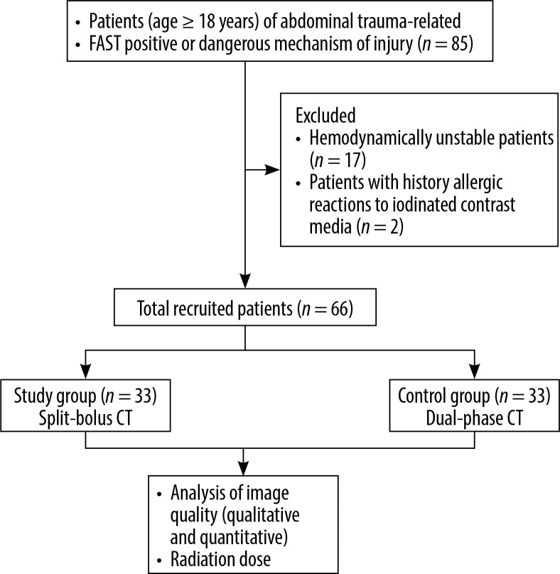
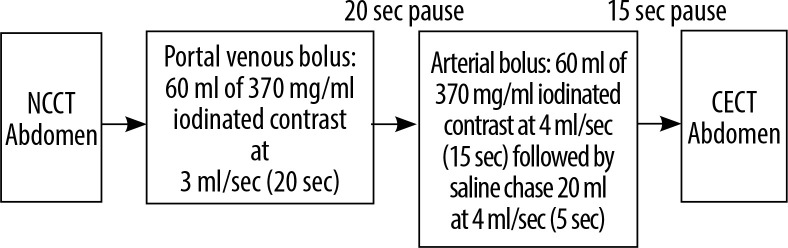
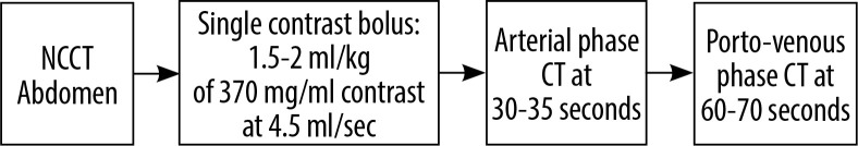
Material and methods: Sixty-six consecutive abdominal trauma patients referred for CT were randomised into 2 groups: the study group (n = 33), scanned using the split-bolus technique; and the control group (n = 33), scanned using the conventional biphasic technique. CT image quality was analysed subjectively by 2 observers based on a 5-point Likert scale. The images were also analysed quantitatively for attenuation values achieved by region of interest (ROI) placements in major arteries, veins, and solid organs. In addition, the radiation dose in terms of the dose length product (DLP) was compared between the 2 groups.
Results: The image quality in both groups ranged from good to excellent in most cases. There was no statistically significant difference in subjective image quality in both the groups as assessed by Likert score. Attenuation values in solid organs and major venous structures were significantly higher in the split-bolus group (p < 0.001). Arterial attenuation values were significantly higher in the control group (p < 0.001), but diagnostic levels were achieved in all patients. There was a reduction of 31.1% in DLP in the split-bolus group.
Conclusions: The split-bolus technique offers comparable image quality and higher solid organ and venous enhancement than conventional biphasic protocol at a reduced radiation dose.

 求助内容:
求助内容: 应助结果提醒方式:


