{"title":"癌细胞来源的外泌体miR-34a通过抑制巨噬细胞的M2极化抑制胰腺腺癌细胞的恶性进展。","authors":"Kui Long, Xiang Kui, Qingbin Zeng, Wenzhi Dong","doi":"10.4081/ejh.2025.4176","DOIUrl":null,"url":null,"abstract":"<p><p>This study aimed to investigate the crosstalk mechanism between pancreatic cancer (PAC) cells and M2 tumor-associated macrophages induced by tumor-derived exosomal miR-34a. MicroRNA and mRNA expression levels were detected using RT-qPCR. Cell Counting Kit-8, wound-healing, transwell assays and flow cytometry were respectively employed to assess cell proliferation, migration, invasion and apoptosis. Enzyme-linked immunosorbent assay was utilized to determine cytokine secretion. Transmission electron microscopy and nanoparticle tracking analyses were performed to detect the exosome morphology and particle size. Phagocytosis of exosomes by macrophages was verified by PKH26 labeling. The effects of exosome-treated macrophages on the epithelial-mesenchymal transition, invasion, and migration of PANC-1 cells were investigated using coculture experiments. The identification of miR-34a's potential targets were determined with TargetScan and validated by a dual-luciferase reporter assay. miR-34a was expressed at low levels in PAC tissues, cells, and exosomes. The overexpression of miR-34a restrains the malignant progression of PANC-1 cells. After miR-34a-overexpressed PANC-1-derived exosomes were phagocytosed by macrophages, the process of M2 polarization in macrophages was obstructed, leading to the suppression of epithelial-mesenchymal transition, migration, and invasion of the cocultured PANC-1 cells. Suppressor of cytokine signaling 3 is a direct target of miR-34a. MiR-34a negatively modulates the suppressor of cytokine signaling 3 to prevent the M2 polarization of macrophages by engaging the Janus kinase/signal transducers and activators of the transcription pathway and influencing the malignancy of PAC cells. miR-34a in cancer cell-derived exosomes inhibits the malignant progression of pancreatic cancer cells by restraining M2 polarization of macrophages.</p>","PeriodicalId":50487,"journal":{"name":"European Journal of Histochemistry","volume":"69 2","pages":""},"PeriodicalIF":2.1000,"publicationDate":"2025-04-07","publicationTypes":"Journal Article","fieldsOfStudy":null,"isOpenAccess":false,"openAccessPdf":"https://www.ncbi.nlm.nih.gov/pmc/articles/PMC12051414/pdf/","citationCount":"0","resultStr":"{\"title\":\"Cancer cell-derived exosomal miR-34a inhibits the malignant progression of pancreatic adenocarcinoma cells by restraining the M2 polarization of macrophages.\",\"authors\":\"Kui Long, Xiang Kui, Qingbin Zeng, Wenzhi Dong\",\"doi\":\"10.4081/ejh.2025.4176\",\"DOIUrl\":null,\"url\":null,\"abstract\":\"<p><p>This study aimed to investigate the crosstalk mechanism between pancreatic cancer (PAC) cells and M2 tumor-associated macrophages induced by tumor-derived exosomal miR-34a. MicroRNA and mRNA expression levels were detected using RT-qPCR. Cell Counting Kit-8, wound-healing, transwell assays and flow cytometry were respectively employed to assess cell proliferation, migration, invasion and apoptosis. Enzyme-linked immunosorbent assay was utilized to determine cytokine secretion. Transmission electron microscopy and nanoparticle tracking analyses were performed to detect the exosome morphology and particle size. Phagocytosis of exosomes by macrophages was verified by PKH26 labeling. The effects of exosome-treated macrophages on the epithelial-mesenchymal transition, invasion, and migration of PANC-1 cells were investigated using coculture experiments. The identification of miR-34a's potential targets were determined with TargetScan and validated by a dual-luciferase reporter assay. miR-34a was expressed at low levels in PAC tissues, cells, and exosomes. The overexpression of miR-34a restrains the malignant progression of PANC-1 cells. After miR-34a-overexpressed PANC-1-derived exosomes were phagocytosed by macrophages, the process of M2 polarization in macrophages was obstructed, leading to the suppression of epithelial-mesenchymal transition, migration, and invasion of the cocultured PANC-1 cells. Suppressor of cytokine signaling 3 is a direct target of miR-34a. MiR-34a negatively modulates the suppressor of cytokine signaling 3 to prevent the M2 polarization of macrophages by engaging the Janus kinase/signal transducers and activators of the transcription pathway and influencing the malignancy of PAC cells. miR-34a in cancer cell-derived exosomes inhibits the malignant progression of pancreatic cancer cells by restraining M2 polarization of macrophages.</p>\",\"PeriodicalId\":50487,\"journal\":{\"name\":\"European Journal of Histochemistry\",\"volume\":\"69 2\",\"pages\":\"\"},\"PeriodicalIF\":2.1000,\"publicationDate\":\"2025-04-07\",\"publicationTypes\":\"Journal Article\",\"fieldsOfStudy\":null,\"isOpenAccess\":false,\"openAccessPdf\":\"https://www.ncbi.nlm.nih.gov/pmc/articles/PMC12051414/pdf/\",\"citationCount\":\"0\",\"resultStr\":null,\"platform\":\"Semanticscholar\",\"paperid\":null,\"PeriodicalName\":\"European Journal of Histochemistry\",\"FirstCategoryId\":\"99\",\"ListUrlMain\":\"https://doi.org/10.4081/ejh.2025.4176\",\"RegionNum\":4,\"RegionCategory\":\"生物学\",\"ArticlePicture\":[],\"TitleCN\":null,\"AbstractTextCN\":null,\"PMCID\":null,\"EPubDate\":\"2025/4/17 0:00:00\",\"PubModel\":\"Epub\",\"JCR\":\"Q4\",\"JCRName\":\"CELL BIOLOGY\",\"Score\":null,\"Total\":0}","platform":"Semanticscholar","paperid":null,"PeriodicalName":"European Journal of Histochemistry","FirstCategoryId":"99","ListUrlMain":"https://doi.org/10.4081/ejh.2025.4176","RegionNum":4,"RegionCategory":"生物学","ArticlePicture":[],"TitleCN":null,"AbstractTextCN":null,"PMCID":null,"EPubDate":"2025/4/17 0:00:00","PubModel":"Epub","JCR":"Q4","JCRName":"CELL BIOLOGY","Score":null,"Total":0}
Cancer cell-derived exosomal miR-34a inhibits the malignant progression of pancreatic adenocarcinoma cells by restraining the M2 polarization of macrophages.
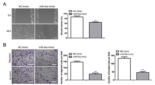
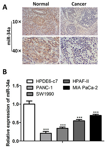
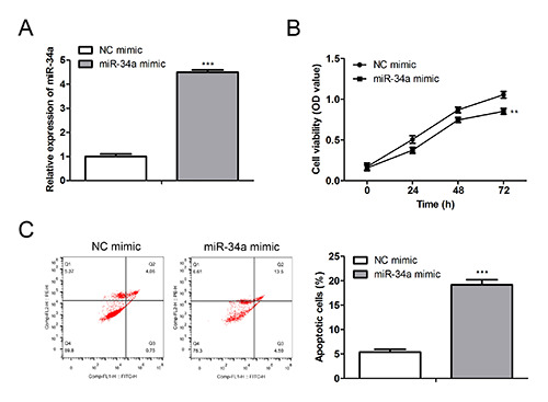
This study aimed to investigate the crosstalk mechanism between pancreatic cancer (PAC) cells and M2 tumor-associated macrophages induced by tumor-derived exosomal miR-34a. MicroRNA and mRNA expression levels were detected using RT-qPCR. Cell Counting Kit-8, wound-healing, transwell assays and flow cytometry were respectively employed to assess cell proliferation, migration, invasion and apoptosis. Enzyme-linked immunosorbent assay was utilized to determine cytokine secretion. Transmission electron microscopy and nanoparticle tracking analyses were performed to detect the exosome morphology and particle size. Phagocytosis of exosomes by macrophages was verified by PKH26 labeling. The effects of exosome-treated macrophages on the epithelial-mesenchymal transition, invasion, and migration of PANC-1 cells were investigated using coculture experiments. The identification of miR-34a's potential targets were determined with TargetScan and validated by a dual-luciferase reporter assay. miR-34a was expressed at low levels in PAC tissues, cells, and exosomes. The overexpression of miR-34a restrains the malignant progression of PANC-1 cells. After miR-34a-overexpressed PANC-1-derived exosomes were phagocytosed by macrophages, the process of M2 polarization in macrophages was obstructed, leading to the suppression of epithelial-mesenchymal transition, migration, and invasion of the cocultured PANC-1 cells. Suppressor of cytokine signaling 3 is a direct target of miR-34a. MiR-34a negatively modulates the suppressor of cytokine signaling 3 to prevent the M2 polarization of macrophages by engaging the Janus kinase/signal transducers and activators of the transcription pathway and influencing the malignancy of PAC cells. miR-34a in cancer cell-derived exosomes inhibits the malignant progression of pancreatic cancer cells by restraining M2 polarization of macrophages.
期刊介绍:
The Journal publishes original papers concerning investigations by histochemical and immunohistochemical methods, and performed with the aid of light, super-resolution and electron microscopy, cytometry and imaging techniques. Coverage extends to:
functional cell and tissue biology in animals and plants;
cell differentiation and death;
cell-cell interaction and molecular trafficking;
biology of cell development and senescence;
nerve and muscle cell biology;
cellular basis of diseases.
The histochemical approach is nowadays essentially aimed at locating molecules in the very place where they exert their biological roles, and at describing dynamically specific chemical activities in living cells. Basic research on cell functional organization is essential for understanding the mechanisms underlying major biological processes such as differentiation, the control of tissue homeostasis, and the regulation of normal and tumor cell growth. Even more than in the past, the European Journal of Histochemistry, as a journal of functional cytology, represents the venue where cell scientists may present and discuss their original results, technical improvements and theories.

 求助内容:
求助内容: 应助结果提醒方式:


