Graham Lloyd-Jones, James Shambrook, Alastair Watson, Anna Freeman, Tom M A Wilkinson
{"title":"胸部计算机断层扫描和x线平片显示COVID-19肺部疾病(肺血管病变)的血管分布和特征。","authors":"Graham Lloyd-Jones, James Shambrook, Alastair Watson, Anna Freeman, Tom M A Wilkinson","doi":"","DOIUrl":null,"url":null,"abstract":"<p><strong>Introduction: </strong>Early in the COVID-19 pandemic, CT was demonstrated as a sensitive tool for diagnosing COVID-19. We undertook a detailed study of CT scans in COVID-19 patients to characterise disease distribution within lung parenchyma, respiratory airways, and pulmonary vasculature, aiming to delineate underlying disease processes.</p><p><strong>Methods: </strong>We characterised acute phase chest CT of 40 participants with COVID-19 from the REACT study, 31 with CT pulmonary angiography (CTPA), 4 with intravenous contrast enhanced CT and 5 with non-intravenous contrast enhanced CT. Participants had neither been vaccinated nor received systemic steroids. We further correlated the distribution of lung parenchymal damage on CT with contemporaneous chest radiographs.</p><p><strong>Results: </strong>Parenchymal lung damage was found in all subjects. However, airways inflammation was present in only 23% (9) and limited to small areas. Notably, vascular abnormalities were dominant and characterised by dilated peripheral pulmonary vessels supplying areas of lung damage in a gravity-dependent distribution bilaterally in 95% (38), basally in 90% (36), peripherally in 92.5% (37), and posteriorly in 90% (36). Macrothrombosis was demonstrated in 23% (7) of CTPAs. Wedge-shaped peripheral lung damage, resembling areas of pulmonary vascular congestion, were distinct in 53% (21) with or without visible macrothrombosis. Pleural effusions were seen in 28% (11). Notably, lung opacification distribution in 98% of the plain radiographs matched distribution on CT (39).</p><p><strong>Conclusion: </strong>Our study frames COVID-19 as a pulmonary vasculopathy rather than a more conventional pneumonia which may be important not only for guiding mechanistic study design but also for the development of novel targeted therapeutics.</p>","PeriodicalId":94250,"journal":{"name":"The Ulster medical journal","volume":"94 1","pages":"4-12"},"PeriodicalIF":0.0000,"publicationDate":"2025-04-01","publicationTypes":"Journal Article","fieldsOfStudy":null,"isOpenAccess":false,"openAccessPdf":"https://www.ncbi.nlm.nih.gov/pmc/articles/PMC12042850/pdf/","citationCount":"0","resultStr":"{\"title\":\"Chest computed tomography and plain radiographs demonstrate vascular distribution and characteristics in COVID-19 lung disease - a pulmonary vasculopathy.\",\"authors\":\"Graham Lloyd-Jones, James Shambrook, Alastair Watson, Anna Freeman, Tom M A Wilkinson\",\"doi\":\"\",\"DOIUrl\":null,\"url\":null,\"abstract\":\"<p><strong>Introduction: </strong>Early in the COVID-19 pandemic, CT was demonstrated as a sensitive tool for diagnosing COVID-19. We undertook a detailed study of CT scans in COVID-19 patients to characterise disease distribution within lung parenchyma, respiratory airways, and pulmonary vasculature, aiming to delineate underlying disease processes.</p><p><strong>Methods: </strong>We characterised acute phase chest CT of 40 participants with COVID-19 from the REACT study, 31 with CT pulmonary angiography (CTPA), 4 with intravenous contrast enhanced CT and 5 with non-intravenous contrast enhanced CT. Participants had neither been vaccinated nor received systemic steroids. We further correlated the distribution of lung parenchymal damage on CT with contemporaneous chest radiographs.</p><p><strong>Results: </strong>Parenchymal lung damage was found in all subjects. However, airways inflammation was present in only 23% (9) and limited to small areas. Notably, vascular abnormalities were dominant and characterised by dilated peripheral pulmonary vessels supplying areas of lung damage in a gravity-dependent distribution bilaterally in 95% (38), basally in 90% (36), peripherally in 92.5% (37), and posteriorly in 90% (36). Macrothrombosis was demonstrated in 23% (7) of CTPAs. Wedge-shaped peripheral lung damage, resembling areas of pulmonary vascular congestion, were distinct in 53% (21) with or without visible macrothrombosis. Pleural effusions were seen in 28% (11). Notably, lung opacification distribution in 98% of the plain radiographs matched distribution on CT (39).</p><p><strong>Conclusion: </strong>Our study frames COVID-19 as a pulmonary vasculopathy rather than a more conventional pneumonia which may be important not only for guiding mechanistic study design but also for the development of novel targeted therapeutics.</p>\",\"PeriodicalId\":94250,\"journal\":{\"name\":\"The Ulster medical journal\",\"volume\":\"94 1\",\"pages\":\"4-12\"},\"PeriodicalIF\":0.0000,\"publicationDate\":\"2025-04-01\",\"publicationTypes\":\"Journal Article\",\"fieldsOfStudy\":null,\"isOpenAccess\":false,\"openAccessPdf\":\"https://www.ncbi.nlm.nih.gov/pmc/articles/PMC12042850/pdf/\",\"citationCount\":\"0\",\"resultStr\":null,\"platform\":\"Semanticscholar\",\"paperid\":null,\"PeriodicalName\":\"The Ulster medical journal\",\"FirstCategoryId\":\"1085\",\"ListUrlMain\":\"\",\"RegionNum\":0,\"RegionCategory\":null,\"ArticlePicture\":[],\"TitleCN\":null,\"AbstractTextCN\":null,\"PMCID\":null,\"EPubDate\":\"2025/4/30 0:00:00\",\"PubModel\":\"Epub\",\"JCR\":\"\",\"JCRName\":\"\",\"Score\":null,\"Total\":0}","platform":"Semanticscholar","paperid":null,"PeriodicalName":"The Ulster medical journal","FirstCategoryId":"1085","ListUrlMain":"","RegionNum":0,"RegionCategory":null,"ArticlePicture":[],"TitleCN":null,"AbstractTextCN":null,"PMCID":null,"EPubDate":"2025/4/30 0:00:00","PubModel":"Epub","JCR":"","JCRName":"","Score":null,"Total":0}
Chest computed tomography and plain radiographs demonstrate vascular distribution and characteristics in COVID-19 lung disease - a pulmonary vasculopathy.
Introduction: Early in the COVID-19 pandemic, CT was demonstrated as a sensitive tool for diagnosing COVID-19. We undertook a detailed study of CT scans in COVID-19 patients to characterise disease distribution within lung parenchyma, respiratory airways, and pulmonary vasculature, aiming to delineate underlying disease processes.
Methods: We characterised acute phase chest CT of 40 participants with COVID-19 from the REACT study, 31 with CT pulmonary angiography (CTPA), 4 with intravenous contrast enhanced CT and 5 with non-intravenous contrast enhanced CT. Participants had neither been vaccinated nor received systemic steroids. We further correlated the distribution of lung parenchymal damage on CT with contemporaneous chest radiographs.
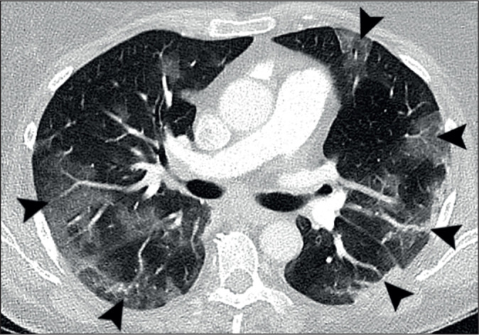
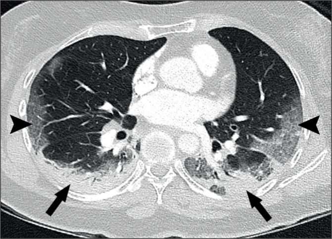
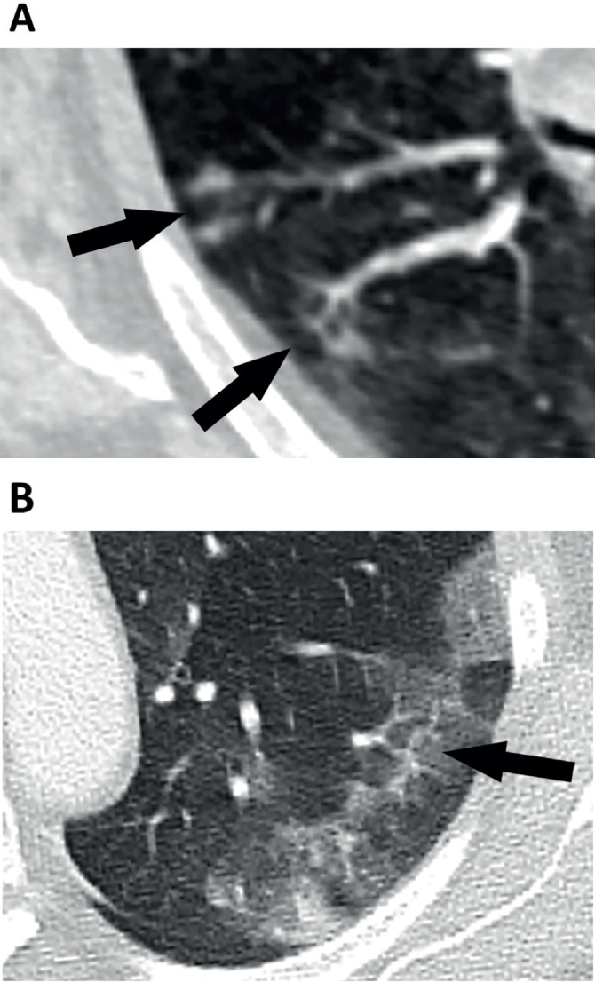
Results: Parenchymal lung damage was found in all subjects. However, airways inflammation was present in only 23% (9) and limited to small areas. Notably, vascular abnormalities were dominant and characterised by dilated peripheral pulmonary vessels supplying areas of lung damage in a gravity-dependent distribution bilaterally in 95% (38), basally in 90% (36), peripherally in 92.5% (37), and posteriorly in 90% (36). Macrothrombosis was demonstrated in 23% (7) of CTPAs. Wedge-shaped peripheral lung damage, resembling areas of pulmonary vascular congestion, were distinct in 53% (21) with or without visible macrothrombosis. Pleural effusions were seen in 28% (11). Notably, lung opacification distribution in 98% of the plain radiographs matched distribution on CT (39).
Conclusion: Our study frames COVID-19 as a pulmonary vasculopathy rather than a more conventional pneumonia which may be important not only for guiding mechanistic study design but also for the development of novel targeted therapeutics.

 求助内容:
求助内容: 应助结果提醒方式:


