{"title":"原发性醛固酮增多症伴68ga - pentxapet /CT阴性和FDG-PET/CT阳性1例左侧肾上腺腺瘤。","authors":"Aamir Nazar, Gaurav Malhotra, Sandip Basu","doi":"10.4103/ijnm.ijnm_150_24","DOIUrl":null,"url":null,"abstract":"<p><p>A 65-year-old male with systemic hypertension, progressively rising aldosterone levels, low plasma renin activity, contrast Magnetic resonance imaging (MRI) described left adrenal lesion isointense to liver parenchyma on T2-weighted images and other features suggestive of left adrenal incidentaloma underwent <sup>68</sup>Ga-Pentixafor Positron emission tomography-computed tomography (PET/CT), which unexpectedly did not show tracer uptake in the known left adrenal adenoma. However, the said nodule showed significant focal tracer uptake on F-18 Fluorodeoxyglucose (FDG)-PET/CT that was done within 2 weeks of <sup>68</sup>Ga-Pentixafor PET/CT study. This unusual finding, which has hitherto been unreported in known patients of primary aldosteronism needs further exploration to determine its clinical significance.</p>","PeriodicalId":45830,"journal":{"name":"Indian Journal of Nuclear Medicine","volume":"39 6","pages":"472-474"},"PeriodicalIF":0.5000,"publicationDate":"2024-11-01","publicationTypes":"Journal Article","fieldsOfStudy":null,"isOpenAccess":false,"openAccessPdf":"https://www.ncbi.nlm.nih.gov/pmc/articles/PMC12020976/pdf/","citationCount":"0","resultStr":"{\"title\":\"An Unusual Case of Primary Aldosteronism with Negative <sup>68</sup>Ga-Pentixafor PET/CT and Positive FDG-PET/CT in a Left Adrenal Adenoma.\",\"authors\":\"Aamir Nazar, Gaurav Malhotra, Sandip Basu\",\"doi\":\"10.4103/ijnm.ijnm_150_24\",\"DOIUrl\":null,\"url\":null,\"abstract\":\"<p><p>A 65-year-old male with systemic hypertension, progressively rising aldosterone levels, low plasma renin activity, contrast Magnetic resonance imaging (MRI) described left adrenal lesion isointense to liver parenchyma on T2-weighted images and other features suggestive of left adrenal incidentaloma underwent <sup>68</sup>Ga-Pentixafor Positron emission tomography-computed tomography (PET/CT), which unexpectedly did not show tracer uptake in the known left adrenal adenoma. However, the said nodule showed significant focal tracer uptake on F-18 Fluorodeoxyglucose (FDG)-PET/CT that was done within 2 weeks of <sup>68</sup>Ga-Pentixafor PET/CT study. This unusual finding, which has hitherto been unreported in known patients of primary aldosteronism needs further exploration to determine its clinical significance.</p>\",\"PeriodicalId\":45830,\"journal\":{\"name\":\"Indian Journal of Nuclear Medicine\",\"volume\":\"39 6\",\"pages\":\"472-474\"},\"PeriodicalIF\":0.5000,\"publicationDate\":\"2024-11-01\",\"publicationTypes\":\"Journal Article\",\"fieldsOfStudy\":null,\"isOpenAccess\":false,\"openAccessPdf\":\"https://www.ncbi.nlm.nih.gov/pmc/articles/PMC12020976/pdf/\",\"citationCount\":\"0\",\"resultStr\":null,\"platform\":\"Semanticscholar\",\"paperid\":null,\"PeriodicalName\":\"Indian Journal of Nuclear Medicine\",\"FirstCategoryId\":\"1085\",\"ListUrlMain\":\"https://doi.org/10.4103/ijnm.ijnm_150_24\",\"RegionNum\":0,\"RegionCategory\":null,\"ArticlePicture\":[],\"TitleCN\":null,\"AbstractTextCN\":null,\"PMCID\":null,\"EPubDate\":\"2025/3/20 0:00:00\",\"PubModel\":\"Epub\",\"JCR\":\"Q4\",\"JCRName\":\"RADIOLOGY, NUCLEAR MEDICINE & MEDICAL IMAGING\",\"Score\":null,\"Total\":0}","platform":"Semanticscholar","paperid":null,"PeriodicalName":"Indian Journal of Nuclear Medicine","FirstCategoryId":"1085","ListUrlMain":"https://doi.org/10.4103/ijnm.ijnm_150_24","RegionNum":0,"RegionCategory":null,"ArticlePicture":[],"TitleCN":null,"AbstractTextCN":null,"PMCID":null,"EPubDate":"2025/3/20 0:00:00","PubModel":"Epub","JCR":"Q4","JCRName":"RADIOLOGY, NUCLEAR MEDICINE & MEDICAL IMAGING","Score":null,"Total":0}
An Unusual Case of Primary Aldosteronism with Negative 68Ga-Pentixafor PET/CT and Positive FDG-PET/CT in a Left Adrenal Adenoma.
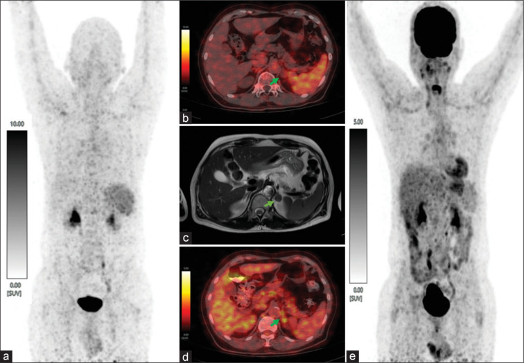
A 65-year-old male with systemic hypertension, progressively rising aldosterone levels, low plasma renin activity, contrast Magnetic resonance imaging (MRI) described left adrenal lesion isointense to liver parenchyma on T2-weighted images and other features suggestive of left adrenal incidentaloma underwent 68Ga-Pentixafor Positron emission tomography-computed tomography (PET/CT), which unexpectedly did not show tracer uptake in the known left adrenal adenoma. However, the said nodule showed significant focal tracer uptake on F-18 Fluorodeoxyglucose (FDG)-PET/CT that was done within 2 weeks of 68Ga-Pentixafor PET/CT study. This unusual finding, which has hitherto been unreported in known patients of primary aldosteronism needs further exploration to determine its clinical significance.

 求助内容:
求助内容: 应助结果提醒方式:


