{"title":"老年猫急性非创伤性腹部假性疝。","authors":"Lauren Stack, QiCai Jason Hoon","doi":"10.1177/20551169251332826","DOIUrl":null,"url":null,"abstract":"<p><strong>Case summary: </strong>A 16-year-old female spayed domestic shorthair cat presented with an acute mass-like outpouching of abdominal viscera unilaterally in the right caudal abdominal ventrum without history of trauma. This abnormality was not identified on prior diagnostic investigations for hyporexia up to 3 weeks prior. A CT examination revealed a protrusion of the viscus through an area of anomalous abdominal wall thinning associated with the discontinuation of the transverse abdominis muscle in this region, rather than a true hernia. Exploratory celiotomy confirmed these findings, with the external rectus sheath and parietal peritoneum remaining intact without an overt hernia ring identified. Abdominal wall augmentation and reconstruction using internal abdominal oblique advancement and fold-over external rectus sheath imbrication resulted in complete resolution without recurrence on subsequent follow-ups. Despite gastrointestinal and muscle biopsies showing no evidence of neoplasia on histopathology, the patient developed an abdominal effusion and was diagnosed with granular lymphocyte lymphoma 4 weeks postoperatively, leading to euthanasia.</p><p><strong>Relevance and novel information: </strong>This case describes a non-traumatic abdominal wall pseudohernia in a geriatric cat, a condition which has not previously been reported. A novel reconstruction technique was used to correct the body wall anomaly, with a good short-term outcome without recurrence.</p>","PeriodicalId":36588,"journal":{"name":"Journal of Feline Medicine and Surgery Open Reports","volume":"11 1","pages":"20551169251332826"},"PeriodicalIF":0.7000,"publicationDate":"2025-05-07","publicationTypes":"Journal Article","fieldsOfStudy":null,"isOpenAccess":false,"openAccessPdf":"https://www.ncbi.nlm.nih.gov/pmc/articles/PMC12062702/pdf/","citationCount":"0","resultStr":"{\"title\":\"Acute non-traumatic abdominal pseudoherniation in a geriatric cat.\",\"authors\":\"Lauren Stack, QiCai Jason Hoon\",\"doi\":\"10.1177/20551169251332826\",\"DOIUrl\":null,\"url\":null,\"abstract\":\"<p><strong>Case summary: </strong>A 16-year-old female spayed domestic shorthair cat presented with an acute mass-like outpouching of abdominal viscera unilaterally in the right caudal abdominal ventrum without history of trauma. This abnormality was not identified on prior diagnostic investigations for hyporexia up to 3 weeks prior. A CT examination revealed a protrusion of the viscus through an area of anomalous abdominal wall thinning associated with the discontinuation of the transverse abdominis muscle in this region, rather than a true hernia. Exploratory celiotomy confirmed these findings, with the external rectus sheath and parietal peritoneum remaining intact without an overt hernia ring identified. Abdominal wall augmentation and reconstruction using internal abdominal oblique advancement and fold-over external rectus sheath imbrication resulted in complete resolution without recurrence on subsequent follow-ups. Despite gastrointestinal and muscle biopsies showing no evidence of neoplasia on histopathology, the patient developed an abdominal effusion and was diagnosed with granular lymphocyte lymphoma 4 weeks postoperatively, leading to euthanasia.</p><p><strong>Relevance and novel information: </strong>This case describes a non-traumatic abdominal wall pseudohernia in a geriatric cat, a condition which has not previously been reported. A novel reconstruction technique was used to correct the body wall anomaly, with a good short-term outcome without recurrence.</p>\",\"PeriodicalId\":36588,\"journal\":{\"name\":\"Journal of Feline Medicine and Surgery Open Reports\",\"volume\":\"11 1\",\"pages\":\"20551169251332826\"},\"PeriodicalIF\":0.7000,\"publicationDate\":\"2025-05-07\",\"publicationTypes\":\"Journal Article\",\"fieldsOfStudy\":null,\"isOpenAccess\":false,\"openAccessPdf\":\"https://www.ncbi.nlm.nih.gov/pmc/articles/PMC12062702/pdf/\",\"citationCount\":\"0\",\"resultStr\":null,\"platform\":\"Semanticscholar\",\"paperid\":null,\"PeriodicalName\":\"Journal of Feline Medicine and Surgery Open Reports\",\"FirstCategoryId\":\"1085\",\"ListUrlMain\":\"https://doi.org/10.1177/20551169251332826\",\"RegionNum\":0,\"RegionCategory\":null,\"ArticlePicture\":[],\"TitleCN\":null,\"AbstractTextCN\":null,\"PMCID\":null,\"EPubDate\":\"2025/1/1 0:00:00\",\"PubModel\":\"eCollection\",\"JCR\":\"Q3\",\"JCRName\":\"VETERINARY SCIENCES\",\"Score\":null,\"Total\":0}","platform":"Semanticscholar","paperid":null,"PeriodicalName":"Journal of Feline Medicine and Surgery Open Reports","FirstCategoryId":"1085","ListUrlMain":"https://doi.org/10.1177/20551169251332826","RegionNum":0,"RegionCategory":null,"ArticlePicture":[],"TitleCN":null,"AbstractTextCN":null,"PMCID":null,"EPubDate":"2025/1/1 0:00:00","PubModel":"eCollection","JCR":"Q3","JCRName":"VETERINARY SCIENCES","Score":null,"Total":0}
Acute non-traumatic abdominal pseudoherniation in a geriatric cat.
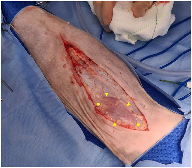
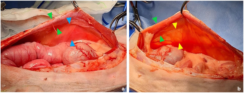
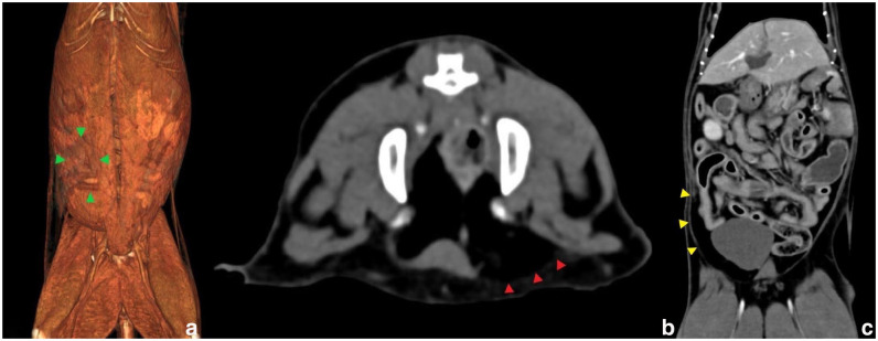
Case summary: A 16-year-old female spayed domestic shorthair cat presented with an acute mass-like outpouching of abdominal viscera unilaterally in the right caudal abdominal ventrum without history of trauma. This abnormality was not identified on prior diagnostic investigations for hyporexia up to 3 weeks prior. A CT examination revealed a protrusion of the viscus through an area of anomalous abdominal wall thinning associated with the discontinuation of the transverse abdominis muscle in this region, rather than a true hernia. Exploratory celiotomy confirmed these findings, with the external rectus sheath and parietal peritoneum remaining intact without an overt hernia ring identified. Abdominal wall augmentation and reconstruction using internal abdominal oblique advancement and fold-over external rectus sheath imbrication resulted in complete resolution without recurrence on subsequent follow-ups. Despite gastrointestinal and muscle biopsies showing no evidence of neoplasia on histopathology, the patient developed an abdominal effusion and was diagnosed with granular lymphocyte lymphoma 4 weeks postoperatively, leading to euthanasia.
Relevance and novel information: This case describes a non-traumatic abdominal wall pseudohernia in a geriatric cat, a condition which has not previously been reported. A novel reconstruction technique was used to correct the body wall anomaly, with a good short-term outcome without recurrence.

 求助内容:
求助内容: 应助结果提醒方式:


