Shamala Sivanandham, Sri Sruthi Preman, Adhithya Baskaran, Kokila Sivakumar
{"title":"脸颊血管平滑肌瘤- 1例报告强调免疫组织化学诊断方法。","authors":"Shamala Sivanandham, Sri Sruthi Preman, Adhithya Baskaran, Kokila Sivakumar","doi":"10.4103/jomfp.jomfp_138_24","DOIUrl":null,"url":null,"abstract":"<p><p>Benign smooth muscle tumours, known as leiomyomas, are comparatively frequent, with uterine cases accounting for 95% incidence. Oral leiomyomas typically appear as asymptomatic, slowly-growing submucosal lumps in the tongue, hard palate, or buccal mucosa. Three histologic kinds of leiomyomas are recognized: epithelioid leiomyoma, solid leiomyoma, and angioleiomyoma. The most prevalent type of leiomyomas affecting the oral cavity are solitary forms called angioleiomyomas, which typically develop in the subcutis. The diagnosis is commonly determined by histopathological and Immunohistochemistry (IHC) procedures. This case reports a 30-year-old female patient with a lesion on her right cheek region. The postsurgical specimen was routinely processed and stained with hematoxylin and eosin staining, and IHC studies confirmed the diagnosis of 'Benign spindle cell neoplasm-Angioleiomyoma'.</p>","PeriodicalId":38846,"journal":{"name":"Journal of Oral and Maxillofacial Pathology","volume":"29 1","pages":"158-162"},"PeriodicalIF":0.0000,"publicationDate":"2025-01-01","publicationTypes":"Journal Article","fieldsOfStudy":null,"isOpenAccess":false,"openAccessPdf":"https://www.ncbi.nlm.nih.gov/pmc/articles/PMC12002582/pdf/","citationCount":"0","resultStr":"{\"title\":\"Angioleiomyoma of cheek - A case report highlighting immunohistochemical diagnostic approach.\",\"authors\":\"Shamala Sivanandham, Sri Sruthi Preman, Adhithya Baskaran, Kokila Sivakumar\",\"doi\":\"10.4103/jomfp.jomfp_138_24\",\"DOIUrl\":null,\"url\":null,\"abstract\":\"<p><p>Benign smooth muscle tumours, known as leiomyomas, are comparatively frequent, with uterine cases accounting for 95% incidence. Oral leiomyomas typically appear as asymptomatic, slowly-growing submucosal lumps in the tongue, hard palate, or buccal mucosa. Three histologic kinds of leiomyomas are recognized: epithelioid leiomyoma, solid leiomyoma, and angioleiomyoma. The most prevalent type of leiomyomas affecting the oral cavity are solitary forms called angioleiomyomas, which typically develop in the subcutis. The diagnosis is commonly determined by histopathological and Immunohistochemistry (IHC) procedures. This case reports a 30-year-old female patient with a lesion on her right cheek region. The postsurgical specimen was routinely processed and stained with hematoxylin and eosin staining, and IHC studies confirmed the diagnosis of 'Benign spindle cell neoplasm-Angioleiomyoma'.</p>\",\"PeriodicalId\":38846,\"journal\":{\"name\":\"Journal of Oral and Maxillofacial Pathology\",\"volume\":\"29 1\",\"pages\":\"158-162\"},\"PeriodicalIF\":0.0000,\"publicationDate\":\"2025-01-01\",\"publicationTypes\":\"Journal Article\",\"fieldsOfStudy\":null,\"isOpenAccess\":false,\"openAccessPdf\":\"https://www.ncbi.nlm.nih.gov/pmc/articles/PMC12002582/pdf/\",\"citationCount\":\"0\",\"resultStr\":null,\"platform\":\"Semanticscholar\",\"paperid\":null,\"PeriodicalName\":\"Journal of Oral and Maxillofacial Pathology\",\"FirstCategoryId\":\"1085\",\"ListUrlMain\":\"https://doi.org/10.4103/jomfp.jomfp_138_24\",\"RegionNum\":0,\"RegionCategory\":null,\"ArticlePicture\":[],\"TitleCN\":null,\"AbstractTextCN\":null,\"PMCID\":null,\"EPubDate\":\"2025/3/28 0:00:00\",\"PubModel\":\"Epub\",\"JCR\":\"Q3\",\"JCRName\":\"Medicine\",\"Score\":null,\"Total\":0}","platform":"Semanticscholar","paperid":null,"PeriodicalName":"Journal of Oral and Maxillofacial Pathology","FirstCategoryId":"1085","ListUrlMain":"https://doi.org/10.4103/jomfp.jomfp_138_24","RegionNum":0,"RegionCategory":null,"ArticlePicture":[],"TitleCN":null,"AbstractTextCN":null,"PMCID":null,"EPubDate":"2025/3/28 0:00:00","PubModel":"Epub","JCR":"Q3","JCRName":"Medicine","Score":null,"Total":0}
Angioleiomyoma of cheek - A case report highlighting immunohistochemical diagnostic approach.
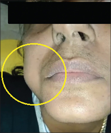
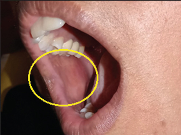
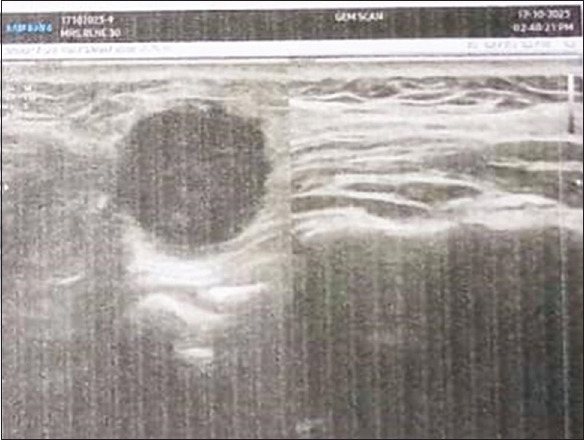
Benign smooth muscle tumours, known as leiomyomas, are comparatively frequent, with uterine cases accounting for 95% incidence. Oral leiomyomas typically appear as asymptomatic, slowly-growing submucosal lumps in the tongue, hard palate, or buccal mucosa. Three histologic kinds of leiomyomas are recognized: epithelioid leiomyoma, solid leiomyoma, and angioleiomyoma. The most prevalent type of leiomyomas affecting the oral cavity are solitary forms called angioleiomyomas, which typically develop in the subcutis. The diagnosis is commonly determined by histopathological and Immunohistochemistry (IHC) procedures. This case reports a 30-year-old female patient with a lesion on her right cheek region. The postsurgical specimen was routinely processed and stained with hematoxylin and eosin staining, and IHC studies confirmed the diagnosis of 'Benign spindle cell neoplasm-Angioleiomyoma'.
期刊介绍:
The journal of Oral and Maxillofacial Pathology [ISSN:print-(0973-029X, online-1998-393X)] is a tri-annual journal published on behalf of “The Indian Association of Oral and Maxillofacial Pathologists” (IAOMP). The publication of JOMFP was started in the year 1993. The journal publishes papers on a wide spectrum of topics associated with the scope of Oral and Maxillofacial Pathology, also, ensuring scientific merit and quality. It is a comprehensive reading material for the professionals who want to upgrade their diagnostic skills in Oral Diseases; allows exposure to newer topics and methods of research in the Oral-facial Tissues and Pathology. New features allow an open minded thinking and approach to various pathologies. It also encourages authors to showcase quality work done by them and to compile relevant cases which are diagnostically challenging. The Journal takes pride in maintaining the quality of articles and photomicrographs.

 求助内容:
求助内容: 应助结果提醒方式:


