Manuel Mazzucchelli, Giuseppe Angelico, Lucia Salvatorelli, Giada Maria Vecchio, Chiara Romano, Paolo Vigneri, Maria Paola Mariani, Gerardo Ferrara, Gaetano Giuseppe Magro
{"title":"腹股沟区脂肪母细胞瘤样肿瘤:黏液样脂肪肉瘤的近似模拟物。","authors":"Manuel Mazzucchelli, Giuseppe Angelico, Lucia Salvatorelli, Giada Maria Vecchio, Chiara Romano, Paolo Vigneri, Maria Paola Mariani, Gerardo Ferrara, Gaetano Giuseppe Magro","doi":"10.32074/1591-951X-964","DOIUrl":null,"url":null,"abstract":"<p><p>Lipoblastoma-like tumor is a rare mesenchymal neoplasm, typically arising in the vulvar region of young women. Although it is considered a benign tumor, rare local recurrences and exceptionally distant metastases have been reported. Histological examination reveals a well-circumscribed tumor with lobulated pattern, composed of a mixture of mature adipocytes, spindle cells and lipoblasts set in abundant myxoid stroma with numerous thin-walled capillary-like vessels. Due to the rarity of this neoplasm and its morphological resemblance with other benign and malignant lipomatous tumors, the diagnosis of <i>lipoblastoma-like tumor</i> is often challenging. Herein, we present a case occurring in the inguinal region of a 28-year-old woman. Histological examination showed a mixture of mature adipocytes, bland-looking spindle cells with fibrillary cytoplasm, and numerous univacuolated lipoblasts set in a prominent myxoid matrix containing numerous thin-walled branching vessels. Immunohistochemically, neoplastic cells showed diffuse immunostaining for CD34 and negativity for α-smooth muscle actin, desmin, Rb1, MDM2 and STAT6. The main differential diagnoses included myxoid liposarcoma, spindle cell lipoma and cellular angiofibroma. FISH was negative for <i>DDIT3</i>; moreover, no evidence of regional gain or loss of <i>RB1</i> was identified by FISH. Based on morphological, immunohistochemical and cytogenetic/molecular findings, a final diagnosis of \"<i>lipoblastoma-like tumor\"</i> of the inguinal region was rendered.</p>","PeriodicalId":45893,"journal":{"name":"PATHOLOGICA","volume":"117 1","pages":"39-44"},"PeriodicalIF":2.9000,"publicationDate":"2025-02-01","publicationTypes":"Journal Article","fieldsOfStudy":null,"isOpenAccess":false,"openAccessPdf":"https://www.ncbi.nlm.nih.gov/pmc/articles/PMC11983084/pdf/","citationCount":"0","resultStr":"{\"title\":\"Lipoblastoma-like tumor of the inguinal region: a close mimicker of myxoid liposarcoma.\",\"authors\":\"Manuel Mazzucchelli, Giuseppe Angelico, Lucia Salvatorelli, Giada Maria Vecchio, Chiara Romano, Paolo Vigneri, Maria Paola Mariani, Gerardo Ferrara, Gaetano Giuseppe Magro\",\"doi\":\"10.32074/1591-951X-964\",\"DOIUrl\":null,\"url\":null,\"abstract\":\"<p><p>Lipoblastoma-like tumor is a rare mesenchymal neoplasm, typically arising in the vulvar region of young women. Although it is considered a benign tumor, rare local recurrences and exceptionally distant metastases have been reported. Histological examination reveals a well-circumscribed tumor with lobulated pattern, composed of a mixture of mature adipocytes, spindle cells and lipoblasts set in abundant myxoid stroma with numerous thin-walled capillary-like vessels. Due to the rarity of this neoplasm and its morphological resemblance with other benign and malignant lipomatous tumors, the diagnosis of <i>lipoblastoma-like tumor</i> is often challenging. Herein, we present a case occurring in the inguinal region of a 28-year-old woman. Histological examination showed a mixture of mature adipocytes, bland-looking spindle cells with fibrillary cytoplasm, and numerous univacuolated lipoblasts set in a prominent myxoid matrix containing numerous thin-walled branching vessels. Immunohistochemically, neoplastic cells showed diffuse immunostaining for CD34 and negativity for α-smooth muscle actin, desmin, Rb1, MDM2 and STAT6. The main differential diagnoses included myxoid liposarcoma, spindle cell lipoma and cellular angiofibroma. FISH was negative for <i>DDIT3</i>; moreover, no evidence of regional gain or loss of <i>RB1</i> was identified by FISH. Based on morphological, immunohistochemical and cytogenetic/molecular findings, a final diagnosis of \\\"<i>lipoblastoma-like tumor\\\"</i> of the inguinal region was rendered.</p>\",\"PeriodicalId\":45893,\"journal\":{\"name\":\"PATHOLOGICA\",\"volume\":\"117 1\",\"pages\":\"39-44\"},\"PeriodicalIF\":2.9000,\"publicationDate\":\"2025-02-01\",\"publicationTypes\":\"Journal Article\",\"fieldsOfStudy\":null,\"isOpenAccess\":false,\"openAccessPdf\":\"https://www.ncbi.nlm.nih.gov/pmc/articles/PMC11983084/pdf/\",\"citationCount\":\"0\",\"resultStr\":null,\"platform\":\"Semanticscholar\",\"paperid\":null,\"PeriodicalName\":\"PATHOLOGICA\",\"FirstCategoryId\":\"1085\",\"ListUrlMain\":\"https://doi.org/10.32074/1591-951X-964\",\"RegionNum\":0,\"RegionCategory\":null,\"ArticlePicture\":[],\"TitleCN\":null,\"AbstractTextCN\":null,\"PMCID\":null,\"EPubDate\":\"\",\"PubModel\":\"\",\"JCR\":\"Q1\",\"JCRName\":\"PATHOLOGY\",\"Score\":null,\"Total\":0}","platform":"Semanticscholar","paperid":null,"PeriodicalName":"PATHOLOGICA","FirstCategoryId":"1085","ListUrlMain":"https://doi.org/10.32074/1591-951X-964","RegionNum":0,"RegionCategory":null,"ArticlePicture":[],"TitleCN":null,"AbstractTextCN":null,"PMCID":null,"EPubDate":"","PubModel":"","JCR":"Q1","JCRName":"PATHOLOGY","Score":null,"Total":0}
Lipoblastoma-like tumor of the inguinal region: a close mimicker of myxoid liposarcoma.
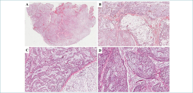
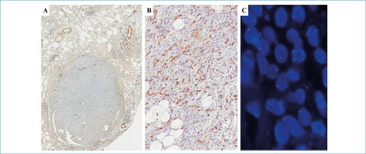
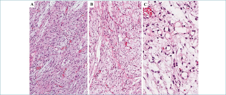
Lipoblastoma-like tumor is a rare mesenchymal neoplasm, typically arising in the vulvar region of young women. Although it is considered a benign tumor, rare local recurrences and exceptionally distant metastases have been reported. Histological examination reveals a well-circumscribed tumor with lobulated pattern, composed of a mixture of mature adipocytes, spindle cells and lipoblasts set in abundant myxoid stroma with numerous thin-walled capillary-like vessels. Due to the rarity of this neoplasm and its morphological resemblance with other benign and malignant lipomatous tumors, the diagnosis of lipoblastoma-like tumor is often challenging. Herein, we present a case occurring in the inguinal region of a 28-year-old woman. Histological examination showed a mixture of mature adipocytes, bland-looking spindle cells with fibrillary cytoplasm, and numerous univacuolated lipoblasts set in a prominent myxoid matrix containing numerous thin-walled branching vessels. Immunohistochemically, neoplastic cells showed diffuse immunostaining for CD34 and negativity for α-smooth muscle actin, desmin, Rb1, MDM2 and STAT6. The main differential diagnoses included myxoid liposarcoma, spindle cell lipoma and cellular angiofibroma. FISH was negative for DDIT3; moreover, no evidence of regional gain or loss of RB1 was identified by FISH. Based on morphological, immunohistochemical and cytogenetic/molecular findings, a final diagnosis of "lipoblastoma-like tumor" of the inguinal region was rendered.

 求助内容:
求助内容: 应助结果提醒方式:


