{"title":"新形成的骨与感染和非感染牙齿是否保留牙槽嵴的实验性种植周炎进展之间的相关性:临床前研究的二次分析","authors":"Jungwoo Jung, Seunghee Lee, Jungwon Lee, Young-Chang Ko, Dongseob Lee, Yang-Jo Seol, Ki-Tae Koo, Yong-Moo Lee","doi":"10.5051/jpis.2402100105","DOIUrl":null,"url":null,"abstract":"<p><strong>Purpose: </strong>We examined the progression of experimental peri-implantitis in sites that underwent either alveolar ridge preservation (ARP) or spontaneous healing (SH), comparing infected teeth (IT) and non-infected teeth (NIT). This investigation is a secondary analysis of a preclinical study aimed at exploring the correlation between newly formed bone and implant stability quotient (ISQ), as well as the association between newly formed bone and the progression of experimental peri-implantitis.</p><p><strong>Methods: </strong>The bilateral mandibular third or fourth premolars of 6 beagle dogs were randomly assigned to 4 groups: IT/SH, IT/ARP, NIT/SH, and NIT/ARP. Following implant placement, core biopsies were retrieved from each site, and the ISQ value was measured. A 3-month period was allowed for peri-implantitis induction, followed by an additional 3 months for the spontaneous progression of peri-implantitis, with radiographs taken at each time point.</p><p><strong>Results: </strong>During the spontaneous progression of peri-implantitis, no statistically significant differences were observed among the groups in terms of mean ISQ values and radiographic marginal bone loss. Similarly, the percentages of bone substitute, newly formed bone, and fibrovascular connective tissue in core biopsies did not differ significantly among the groups. Linear regression analysis revealed no significant linear correlation between newly formed bone and ISQ in any group (<i>P</i>>0.05). However, a weak linear correlation between newly formed bone and marginal bone loss during the spontaneous progression of peri-implantitis was noted in the IT/SH group alone (<i>P</i>=0.036).</p><p><strong>Conclusions: </strong>Within the limitations of this study, we were unable to demonstrate that ARP could improve newly formed bone or primary implant stability. Furthermore, neither ARP nor SH significantly influenced the spontaneous progression of experimental peri-implantitis.</p>","PeriodicalId":48795,"journal":{"name":"Journal of Periodontal and Implant Science","volume":"55 2","pages":"139-152"},"PeriodicalIF":3.2000,"publicationDate":"2025-04-01","publicationTypes":"Journal Article","fieldsOfStudy":null,"isOpenAccess":false,"openAccessPdf":"https://www.ncbi.nlm.nih.gov/pmc/articles/PMC12056239/pdf/","citationCount":"0","resultStr":"{\"title\":\"Correlation between newly formed bone and the progression of experimental peri-implantitis with or without alveolar ridge preservation in infected and non-infected teeth: a secondary analysis of a preclinical study.\",\"authors\":\"Jungwoo Jung, Seunghee Lee, Jungwon Lee, Young-Chang Ko, Dongseob Lee, Yang-Jo Seol, Ki-Tae Koo, Yong-Moo Lee\",\"doi\":\"10.5051/jpis.2402100105\",\"DOIUrl\":null,\"url\":null,\"abstract\":\"<p><strong>Purpose: </strong>We examined the progression of experimental peri-implantitis in sites that underwent either alveolar ridge preservation (ARP) or spontaneous healing (SH), comparing infected teeth (IT) and non-infected teeth (NIT). This investigation is a secondary analysis of a preclinical study aimed at exploring the correlation between newly formed bone and implant stability quotient (ISQ), as well as the association between newly formed bone and the progression of experimental peri-implantitis.</p><p><strong>Methods: </strong>The bilateral mandibular third or fourth premolars of 6 beagle dogs were randomly assigned to 4 groups: IT/SH, IT/ARP, NIT/SH, and NIT/ARP. Following implant placement, core biopsies were retrieved from each site, and the ISQ value was measured. A 3-month period was allowed for peri-implantitis induction, followed by an additional 3 months for the spontaneous progression of peri-implantitis, with radiographs taken at each time point.</p><p><strong>Results: </strong>During the spontaneous progression of peri-implantitis, no statistically significant differences were observed among the groups in terms of mean ISQ values and radiographic marginal bone loss. Similarly, the percentages of bone substitute, newly formed bone, and fibrovascular connective tissue in core biopsies did not differ significantly among the groups. Linear regression analysis revealed no significant linear correlation between newly formed bone and ISQ in any group (<i>P</i>>0.05). However, a weak linear correlation between newly formed bone and marginal bone loss during the spontaneous progression of peri-implantitis was noted in the IT/SH group alone (<i>P</i>=0.036).</p><p><strong>Conclusions: </strong>Within the limitations of this study, we were unable to demonstrate that ARP could improve newly formed bone or primary implant stability. Furthermore, neither ARP nor SH significantly influenced the spontaneous progression of experimental peri-implantitis.</p>\",\"PeriodicalId\":48795,\"journal\":{\"name\":\"Journal of Periodontal and Implant Science\",\"volume\":\"55 2\",\"pages\":\"139-152\"},\"PeriodicalIF\":3.2000,\"publicationDate\":\"2025-04-01\",\"publicationTypes\":\"Journal Article\",\"fieldsOfStudy\":null,\"isOpenAccess\":false,\"openAccessPdf\":\"https://www.ncbi.nlm.nih.gov/pmc/articles/PMC12056239/pdf/\",\"citationCount\":\"0\",\"resultStr\":null,\"platform\":\"Semanticscholar\",\"paperid\":null,\"PeriodicalName\":\"Journal of Periodontal and Implant Science\",\"FirstCategoryId\":\"3\",\"ListUrlMain\":\"https://doi.org/10.5051/jpis.2402100105\",\"RegionNum\":4,\"RegionCategory\":\"医学\",\"ArticlePicture\":[],\"TitleCN\":null,\"AbstractTextCN\":null,\"PMCID\":null,\"EPubDate\":\"\",\"PubModel\":\"\",\"JCR\":\"Q2\",\"JCRName\":\"DENTISTRY, ORAL SURGERY & MEDICINE\",\"Score\":null,\"Total\":0}","platform":"Semanticscholar","paperid":null,"PeriodicalName":"Journal of Periodontal and Implant Science","FirstCategoryId":"3","ListUrlMain":"https://doi.org/10.5051/jpis.2402100105","RegionNum":4,"RegionCategory":"医学","ArticlePicture":[],"TitleCN":null,"AbstractTextCN":null,"PMCID":null,"EPubDate":"","PubModel":"","JCR":"Q2","JCRName":"DENTISTRY, ORAL SURGERY & MEDICINE","Score":null,"Total":0}
Correlation between newly formed bone and the progression of experimental peri-implantitis with or without alveolar ridge preservation in infected and non-infected teeth: a secondary analysis of a preclinical study.
Purpose: We examined the progression of experimental peri-implantitis in sites that underwent either alveolar ridge preservation (ARP) or spontaneous healing (SH), comparing infected teeth (IT) and non-infected teeth (NIT). This investigation is a secondary analysis of a preclinical study aimed at exploring the correlation between newly formed bone and implant stability quotient (ISQ), as well as the association between newly formed bone and the progression of experimental peri-implantitis.
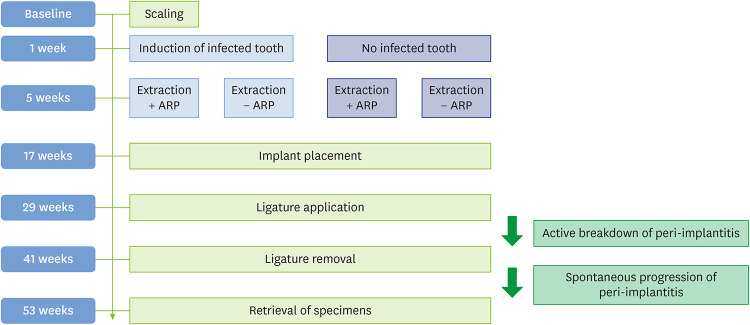
Methods: The bilateral mandibular third or fourth premolars of 6 beagle dogs were randomly assigned to 4 groups: IT/SH, IT/ARP, NIT/SH, and NIT/ARP. Following implant placement, core biopsies were retrieved from each site, and the ISQ value was measured. A 3-month period was allowed for peri-implantitis induction, followed by an additional 3 months for the spontaneous progression of peri-implantitis, with radiographs taken at each time point.
Results: During the spontaneous progression of peri-implantitis, no statistically significant differences were observed among the groups in terms of mean ISQ values and radiographic marginal bone loss. Similarly, the percentages of bone substitute, newly formed bone, and fibrovascular connective tissue in core biopsies did not differ significantly among the groups. Linear regression analysis revealed no significant linear correlation between newly formed bone and ISQ in any group (P>0.05). However, a weak linear correlation between newly formed bone and marginal bone loss during the spontaneous progression of peri-implantitis was noted in the IT/SH group alone (P=0.036).
Conclusions: Within the limitations of this study, we were unable to demonstrate that ARP could improve newly formed bone or primary implant stability. Furthermore, neither ARP nor SH significantly influenced the spontaneous progression of experimental peri-implantitis.
期刊介绍:
Journal of Periodontal & Implant Science (JPIS) is a peer-reviewed and open-access journal providing up-to-date information relevant to professionalism of periodontology and dental implantology. JPIS is dedicated to global and extensive publication which includes evidence-based original articles, and fundamental reviews in order to cover a variety of interests in the field of periodontal as well as implant science.


 求助内容:
求助内容: 应助结果提醒方式:


