Tathagata Bhattacharjee, Supratim Ghosh, Dipto De, Pratik Sarkar, Jay Gopal Ray
{"title":"晚期口腔黏膜下纤维化与恶性口腔黏膜下纤维化的血管形态学比较分析。","authors":"Tathagata Bhattacharjee, Supratim Ghosh, Dipto De, Pratik Sarkar, Jay Gopal Ray","doi":"10.4103/jomfp.jomfp_89_24","DOIUrl":null,"url":null,"abstract":"<p><strong>Introduction: </strong>Oral submucous fibrosis (OSF) is a potentially malignant disorder of oral cavity. The vascularity of submucosa in this disease can predict its malignant transformation.</p><p><strong>Aim: </strong>In this study, attempts have been made to investigate different parameters of vascularity, such as mean vascular area, luminal diameter etc., in advanced OSF and make a comparison with malignancy associated with OSF.</p><p><strong>Materials and methods: </strong>Incisional biopsy was taken from clinically diagnosed cases of advanced OSF and malignancy associated with OSF. Sections were prepared and stained with haematoxylin and eosin for histomorphometric analysis using a research microscope.</p><p><strong>Results: </strong>A statistically significant increase in mean vascular area and luminal diameter of blood vessels was noticed in malignancy associated with OSF compared to advanced OSF.</p><p><strong>Conclusion: </strong>To support the nutritional demand of carcinogenesis, increased blood supply is required; hence blood supply increased in the form of budding blood vessels or enlarged dilated blood vessels. These changes can be used as a predictive marker for this transformation.</p>","PeriodicalId":38846,"journal":{"name":"Journal of Oral and Maxillofacial Pathology","volume":"29 1","pages":"12-17"},"PeriodicalIF":0.0000,"publicationDate":"2025-01-01","publicationTypes":"Journal Article","fieldsOfStudy":null,"isOpenAccess":false,"openAccessPdf":"https://www.ncbi.nlm.nih.gov/pmc/articles/PMC12002574/pdf/","citationCount":"0","resultStr":"{\"title\":\"Comparative analysis of blood vessel morphometry in advanced oral submucous fibrosis and malignancy associated with oral submucous fibrosis.\",\"authors\":\"Tathagata Bhattacharjee, Supratim Ghosh, Dipto De, Pratik Sarkar, Jay Gopal Ray\",\"doi\":\"10.4103/jomfp.jomfp_89_24\",\"DOIUrl\":null,\"url\":null,\"abstract\":\"<p><strong>Introduction: </strong>Oral submucous fibrosis (OSF) is a potentially malignant disorder of oral cavity. The vascularity of submucosa in this disease can predict its malignant transformation.</p><p><strong>Aim: </strong>In this study, attempts have been made to investigate different parameters of vascularity, such as mean vascular area, luminal diameter etc., in advanced OSF and make a comparison with malignancy associated with OSF.</p><p><strong>Materials and methods: </strong>Incisional biopsy was taken from clinically diagnosed cases of advanced OSF and malignancy associated with OSF. Sections were prepared and stained with haematoxylin and eosin for histomorphometric analysis using a research microscope.</p><p><strong>Results: </strong>A statistically significant increase in mean vascular area and luminal diameter of blood vessels was noticed in malignancy associated with OSF compared to advanced OSF.</p><p><strong>Conclusion: </strong>To support the nutritional demand of carcinogenesis, increased blood supply is required; hence blood supply increased in the form of budding blood vessels or enlarged dilated blood vessels. These changes can be used as a predictive marker for this transformation.</p>\",\"PeriodicalId\":38846,\"journal\":{\"name\":\"Journal of Oral and Maxillofacial Pathology\",\"volume\":\"29 1\",\"pages\":\"12-17\"},\"PeriodicalIF\":0.0000,\"publicationDate\":\"2025-01-01\",\"publicationTypes\":\"Journal Article\",\"fieldsOfStudy\":null,\"isOpenAccess\":false,\"openAccessPdf\":\"https://www.ncbi.nlm.nih.gov/pmc/articles/PMC12002574/pdf/\",\"citationCount\":\"0\",\"resultStr\":null,\"platform\":\"Semanticscholar\",\"paperid\":null,\"PeriodicalName\":\"Journal of Oral and Maxillofacial Pathology\",\"FirstCategoryId\":\"1085\",\"ListUrlMain\":\"https://doi.org/10.4103/jomfp.jomfp_89_24\",\"RegionNum\":0,\"RegionCategory\":null,\"ArticlePicture\":[],\"TitleCN\":null,\"AbstractTextCN\":null,\"PMCID\":null,\"EPubDate\":\"2025/3/28 0:00:00\",\"PubModel\":\"Epub\",\"JCR\":\"Q3\",\"JCRName\":\"Medicine\",\"Score\":null,\"Total\":0}","platform":"Semanticscholar","paperid":null,"PeriodicalName":"Journal of Oral and Maxillofacial Pathology","FirstCategoryId":"1085","ListUrlMain":"https://doi.org/10.4103/jomfp.jomfp_89_24","RegionNum":0,"RegionCategory":null,"ArticlePicture":[],"TitleCN":null,"AbstractTextCN":null,"PMCID":null,"EPubDate":"2025/3/28 0:00:00","PubModel":"Epub","JCR":"Q3","JCRName":"Medicine","Score":null,"Total":0}
Comparative analysis of blood vessel morphometry in advanced oral submucous fibrosis and malignancy associated with oral submucous fibrosis.
Introduction: Oral submucous fibrosis (OSF) is a potentially malignant disorder of oral cavity. The vascularity of submucosa in this disease can predict its malignant transformation.
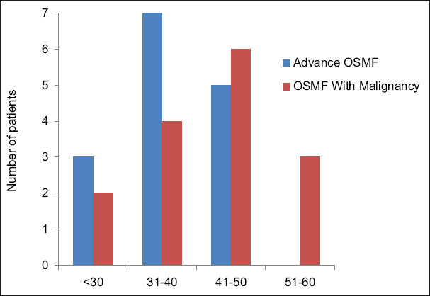
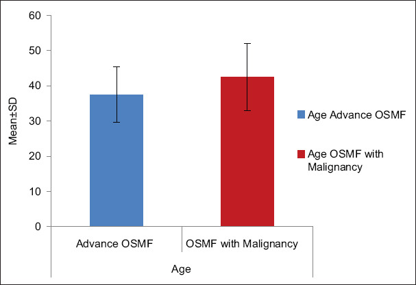
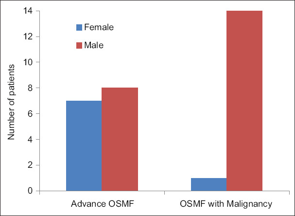
Aim: In this study, attempts have been made to investigate different parameters of vascularity, such as mean vascular area, luminal diameter etc., in advanced OSF and make a comparison with malignancy associated with OSF.
Materials and methods: Incisional biopsy was taken from clinically diagnosed cases of advanced OSF and malignancy associated with OSF. Sections were prepared and stained with haematoxylin and eosin for histomorphometric analysis using a research microscope.
Results: A statistically significant increase in mean vascular area and luminal diameter of blood vessels was noticed in malignancy associated with OSF compared to advanced OSF.
Conclusion: To support the nutritional demand of carcinogenesis, increased blood supply is required; hence blood supply increased in the form of budding blood vessels or enlarged dilated blood vessels. These changes can be used as a predictive marker for this transformation.
期刊介绍:
The journal of Oral and Maxillofacial Pathology [ISSN:print-(0973-029X, online-1998-393X)] is a tri-annual journal published on behalf of “The Indian Association of Oral and Maxillofacial Pathologists” (IAOMP). The publication of JOMFP was started in the year 1993. The journal publishes papers on a wide spectrum of topics associated with the scope of Oral and Maxillofacial Pathology, also, ensuring scientific merit and quality. It is a comprehensive reading material for the professionals who want to upgrade their diagnostic skills in Oral Diseases; allows exposure to newer topics and methods of research in the Oral-facial Tissues and Pathology. New features allow an open minded thinking and approach to various pathologies. It also encourages authors to showcase quality work done by them and to compile relevant cases which are diagnostically challenging. The Journal takes pride in maintaining the quality of articles and photomicrographs.

 求助内容:
求助内容: 应助结果提醒方式:


