Hyeeun Jo, Sang-Kwon Lee, Sooyoung Choi, Miori Kishimoto, Kija Lee
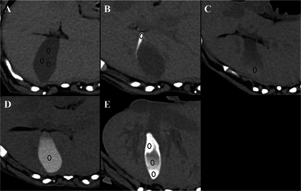
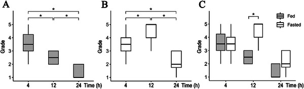
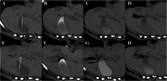
{"title":"在健康犬的延迟计算机断层扫描上可以观察到代入造影剂排泄导致的胆囊和小肠腔混浊。","authors":"Hyeeun Jo, Sang-Kwon Lee, Sooyoung Choi, Miori Kishimoto, Kija Lee","doi":"10.1111/vru.70033","DOIUrl":null,"url":null,"abstract":"<p><p>Vicarious excretion is a nonrenal pathway of excretion for intravenously injected iodinated contrast media, with a limited understanding of its influencing factors and imaging features. In this prospective pilot study, gallbladder opacification (GBO) and small intestinal luminal opacification (SILO) were assessed to identify vicarious excretion patterns following intravenous contrast media administration for CT in clinically healthy dogs. Eight beagles were studied using a crossover method, divided into fed and fasted groups. The fed group was fed at 5 and 13 h after the first CT scan, while the fasted group was fed only at 13 h. Noncontrast and postcontrast CT scans were performed at 90 s, 10 min, 1 h, 4 h, 12 h, and 24 h after iohexol injection. The GBO was subjectively scored from grade 0 to 5 based on the attenuation value and area of opacification. The SILO was evaluated subjectively based on contrast intensity (weak or marked) and distribution. The GBO was observed from 1 h after contrast injection. Significant differences were noted in median GBO scores within the groups at 4, 12, and 24 h on Friedman's test followed by the post hoc Wilcoxon signed-rank test. The scores were significantly higher in the fasted group at 12 h on the Wilcoxon signed-rank test. The SILO occurred 10 min after contrast administration, with various distributions. In conclusion, GBO and SILO can be observed during delayed CT phases, and fasting increases the intensity and duration of GBO in clinically healthy dogs. These findings should not be interpreted as pathological changes.</p>","PeriodicalId":23581,"journal":{"name":"Veterinary Radiology & Ultrasound","volume":"66 3","pages":"e70033"},"PeriodicalIF":1.5000,"publicationDate":"2025-05-01","publicationTypes":"Journal Article","fieldsOfStudy":null,"isOpenAccess":false,"openAccessPdf":"https://www.ncbi.nlm.nih.gov/pmc/articles/PMC12046108/pdf/","citationCount":"0","resultStr":"{\"title\":\"Gallbladder and Small Intestinal Luminal Opacification by Vicarious Contrast Medium Excretion Can be Observed on Delayed Computed Tomography in Healthy Dogs.\",\"authors\":\"Hyeeun Jo, Sang-Kwon Lee, Sooyoung Choi, Miori Kishimoto, Kija Lee\",\"doi\":\"10.1111/vru.70033\",\"DOIUrl\":null,\"url\":null,\"abstract\":\"<p><p>Vicarious excretion is a nonrenal pathway of excretion for intravenously injected iodinated contrast media, with a limited understanding of its influencing factors and imaging features. In this prospective pilot study, gallbladder opacification (GBO) and small intestinal luminal opacification (SILO) were assessed to identify vicarious excretion patterns following intravenous contrast media administration for CT in clinically healthy dogs. Eight beagles were studied using a crossover method, divided into fed and fasted groups. The fed group was fed at 5 and 13 h after the first CT scan, while the fasted group was fed only at 13 h. Noncontrast and postcontrast CT scans were performed at 90 s, 10 min, 1 h, 4 h, 12 h, and 24 h after iohexol injection. The GBO was subjectively scored from grade 0 to 5 based on the attenuation value and area of opacification. The SILO was evaluated subjectively based on contrast intensity (weak or marked) and distribution. The GBO was observed from 1 h after contrast injection. Significant differences were noted in median GBO scores within the groups at 4, 12, and 24 h on Friedman's test followed by the post hoc Wilcoxon signed-rank test. The scores were significantly higher in the fasted group at 12 h on the Wilcoxon signed-rank test. The SILO occurred 10 min after contrast administration, with various distributions. In conclusion, GBO and SILO can be observed during delayed CT phases, and fasting increases the intensity and duration of GBO in clinically healthy dogs. These findings should not be interpreted as pathological changes.</p>\",\"PeriodicalId\":23581,\"journal\":{\"name\":\"Veterinary Radiology & Ultrasound\",\"volume\":\"66 3\",\"pages\":\"e70033\"},\"PeriodicalIF\":1.5000,\"publicationDate\":\"2025-05-01\",\"publicationTypes\":\"Journal Article\",\"fieldsOfStudy\":null,\"isOpenAccess\":false,\"openAccessPdf\":\"https://www.ncbi.nlm.nih.gov/pmc/articles/PMC12046108/pdf/\",\"citationCount\":\"0\",\"resultStr\":null,\"platform\":\"Semanticscholar\",\"paperid\":null,\"PeriodicalName\":\"Veterinary Radiology & Ultrasound\",\"FirstCategoryId\":\"97\",\"ListUrlMain\":\"https://doi.org/10.1111/vru.70033\",\"RegionNum\":2,\"RegionCategory\":\"农林科学\",\"ArticlePicture\":[],\"TitleCN\":null,\"AbstractTextCN\":null,\"PMCID\":null,\"EPubDate\":\"\",\"PubModel\":\"\",\"JCR\":\"Q2\",\"JCRName\":\"VETERINARY SCIENCES\",\"Score\":null,\"Total\":0}","platform":"Semanticscholar","paperid":null,"PeriodicalName":"Veterinary Radiology & Ultrasound","FirstCategoryId":"97","ListUrlMain":"https://doi.org/10.1111/vru.70033","RegionNum":2,"RegionCategory":"农林科学","ArticlePicture":[],"TitleCN":null,"AbstractTextCN":null,"PMCID":null,"EPubDate":"","PubModel":"","JCR":"Q2","JCRName":"VETERINARY SCIENCES","Score":null,"Total":0}
Gallbladder and Small Intestinal Luminal Opacification by Vicarious Contrast Medium Excretion Can be Observed on Delayed Computed Tomography in Healthy Dogs.
Vicarious excretion is a nonrenal pathway of excretion for intravenously injected iodinated contrast media, with a limited understanding of its influencing factors and imaging features. In this prospective pilot study, gallbladder opacification (GBO) and small intestinal luminal opacification (SILO) were assessed to identify vicarious excretion patterns following intravenous contrast media administration for CT in clinically healthy dogs. Eight beagles were studied using a crossover method, divided into fed and fasted groups. The fed group was fed at 5 and 13 h after the first CT scan, while the fasted group was fed only at 13 h. Noncontrast and postcontrast CT scans were performed at 90 s, 10 min, 1 h, 4 h, 12 h, and 24 h after iohexol injection. The GBO was subjectively scored from grade 0 to 5 based on the attenuation value and area of opacification. The SILO was evaluated subjectively based on contrast intensity (weak or marked) and distribution. The GBO was observed from 1 h after contrast injection. Significant differences were noted in median GBO scores within the groups at 4, 12, and 24 h on Friedman's test followed by the post hoc Wilcoxon signed-rank test. The scores were significantly higher in the fasted group at 12 h on the Wilcoxon signed-rank test. The SILO occurred 10 min after contrast administration, with various distributions. In conclusion, GBO and SILO can be observed during delayed CT phases, and fasting increases the intensity and duration of GBO in clinically healthy dogs. These findings should not be interpreted as pathological changes.
期刊介绍:
Veterinary Radiology & Ultrasound is a bimonthly, international, peer-reviewed, research journal devoted to the fields of veterinary diagnostic imaging and radiation oncology. Established in 1958, it is owned by the American College of Veterinary Radiology and is also the official journal for six affiliate veterinary organizations. Veterinary Radiology & Ultrasound is represented on the International Committee of Medical Journal Editors, World Association of Medical Editors, and Committee on Publication Ethics.
The mission of Veterinary Radiology & Ultrasound is to serve as a leading resource for high quality articles that advance scientific knowledge and standards of clinical practice in the areas of veterinary diagnostic radiology, computed tomography, magnetic resonance imaging, ultrasonography, nuclear imaging, radiation oncology, and interventional radiology. Manuscript types include original investigations, imaging diagnosis reports, review articles, editorials and letters to the Editor. Acceptance criteria include originality, significance, quality, reader interest, composition and adherence to author guidelines.

 求助内容:
求助内容: 应助结果提醒方式:


