Burcu Gül, Bora Korkmazer, Ahmet Kürşat Karaman, Esra Koçhan Kizilkiliç, Mahmut Esat Aykan, Çiğdem Özkara, Nil Çomunoğlu, Cihan Işler, Serdar Arslan, Osman Kizilkiliç
{"title":"多结节和空泡神经元肿瘤与胚胎发育异常神经上皮肿瘤的MRI鉴别。","authors":"Burcu Gül, Bora Korkmazer, Ahmet Kürşat Karaman, Esra Koçhan Kizilkiliç, Mahmut Esat Aykan, Çiğdem Özkara, Nil Çomunoğlu, Cihan Işler, Serdar Arslan, Osman Kizilkiliç","doi":"10.55730/1300-0144.5988","DOIUrl":null,"url":null,"abstract":"<p><strong>Background/aim: </strong>To compare the MRI findings and clinical features of multinodular and vacuolating neuronal tumor (MVNT) and dysembryoplastic neuroepithelial tumor (DNET), and reveal the distinguishing features of these tumors from each other.</p><p><strong>Materials and methods: </strong>Patients with a suspected magnetic resonance imaging (MRI)-based diagnosis of MVNT between 2018 and 2022 were collected from the hospital database. In addition, patients diagnosed with DNET on histopathological examination and who had MRIs in the same time period were included in the study. The MRI findings and clinical features were evaluated for each patient.</p><p><strong>Results: </strong>There were 21 patients in the MVNT group and 20 patients in the DNET group. Headache was the most common symptom in patients with MVNTs (61.9%), whereas seizures were more prevalent in those with DNETs (70%). The most frequent locations for the MVNTs were the frontal and parietal lobes (66.6%), while DNETs were most commonly located in the temporal lobe (60%). All the MVNTs were hyperintense in both fluid-attenuated inversion recovery (FLAIR) and T2-weighted imaging (T2WI). All the DNETs were hyperintense on T2WI. However, on FLAIR, seven (35%) of the DNET lesions were hyperintense, while the remaining 13 lesions showed mixed signal intensity forming a bubbly appearance. Moreover, 20 of 21 (95.23%) MVNTs were hyperintense on diffusion-weighted imaging (DWI) (b800), with no apparent diffusion coefficient hypointensity in the lesions. None of the DNETs showed hyperintensity on DWI.</p><p><strong>Conclusion: </strong>MRI findings, particularly those observed on FLAIR and DWI, may be helpful for distinguishing between MVNTs and DNETs, especially in cases where the differential diagnosis is challenging.</p>","PeriodicalId":23361,"journal":{"name":"Turkish Journal of Medical Sciences","volume":"55 2","pages":"443-450"},"PeriodicalIF":1.0000,"publicationDate":"2024-12-30","publicationTypes":"Journal Article","fieldsOfStudy":null,"isOpenAccess":false,"openAccessPdf":"https://www.ncbi.nlm.nih.gov/pmc/articles/PMC12058023/pdf/","citationCount":"0","resultStr":"{\"title\":\"Differentiation of multinodular and vacuolating neuronal tumor and dysembryoplastic neuroepithelial tumor based on MRI.\",\"authors\":\"Burcu Gül, Bora Korkmazer, Ahmet Kürşat Karaman, Esra Koçhan Kizilkiliç, Mahmut Esat Aykan, Çiğdem Özkara, Nil Çomunoğlu, Cihan Işler, Serdar Arslan, Osman Kizilkiliç\",\"doi\":\"10.55730/1300-0144.5988\",\"DOIUrl\":null,\"url\":null,\"abstract\":\"<p><strong>Background/aim: </strong>To compare the MRI findings and clinical features of multinodular and vacuolating neuronal tumor (MVNT) and dysembryoplastic neuroepithelial tumor (DNET), and reveal the distinguishing features of these tumors from each other.</p><p><strong>Materials and methods: </strong>Patients with a suspected magnetic resonance imaging (MRI)-based diagnosis of MVNT between 2018 and 2022 were collected from the hospital database. In addition, patients diagnosed with DNET on histopathological examination and who had MRIs in the same time period were included in the study. The MRI findings and clinical features were evaluated for each patient.</p><p><strong>Results: </strong>There were 21 patients in the MVNT group and 20 patients in the DNET group. Headache was the most common symptom in patients with MVNTs (61.9%), whereas seizures were more prevalent in those with DNETs (70%). The most frequent locations for the MVNTs were the frontal and parietal lobes (66.6%), while DNETs were most commonly located in the temporal lobe (60%). All the MVNTs were hyperintense in both fluid-attenuated inversion recovery (FLAIR) and T2-weighted imaging (T2WI). All the DNETs were hyperintense on T2WI. However, on FLAIR, seven (35%) of the DNET lesions were hyperintense, while the remaining 13 lesions showed mixed signal intensity forming a bubbly appearance. Moreover, 20 of 21 (95.23%) MVNTs were hyperintense on diffusion-weighted imaging (DWI) (b800), with no apparent diffusion coefficient hypointensity in the lesions. None of the DNETs showed hyperintensity on DWI.</p><p><strong>Conclusion: </strong>MRI findings, particularly those observed on FLAIR and DWI, may be helpful for distinguishing between MVNTs and DNETs, especially in cases where the differential diagnosis is challenging.</p>\",\"PeriodicalId\":23361,\"journal\":{\"name\":\"Turkish Journal of Medical Sciences\",\"volume\":\"55 2\",\"pages\":\"443-450\"},\"PeriodicalIF\":1.0000,\"publicationDate\":\"2024-12-30\",\"publicationTypes\":\"Journal Article\",\"fieldsOfStudy\":null,\"isOpenAccess\":false,\"openAccessPdf\":\"https://www.ncbi.nlm.nih.gov/pmc/articles/PMC12058023/pdf/\",\"citationCount\":\"0\",\"resultStr\":null,\"platform\":\"Semanticscholar\",\"paperid\":null,\"PeriodicalName\":\"Turkish Journal of Medical Sciences\",\"FirstCategoryId\":\"3\",\"ListUrlMain\":\"https://doi.org/10.55730/1300-0144.5988\",\"RegionNum\":4,\"RegionCategory\":\"医学\",\"ArticlePicture\":[],\"TitleCN\":null,\"AbstractTextCN\":null,\"PMCID\":null,\"EPubDate\":\"2025/1/1 0:00:00\",\"PubModel\":\"eCollection\",\"JCR\":\"Q2\",\"JCRName\":\"MEDICINE, GENERAL & INTERNAL\",\"Score\":null,\"Total\":0}","platform":"Semanticscholar","paperid":null,"PeriodicalName":"Turkish Journal of Medical Sciences","FirstCategoryId":"3","ListUrlMain":"https://doi.org/10.55730/1300-0144.5988","RegionNum":4,"RegionCategory":"医学","ArticlePicture":[],"TitleCN":null,"AbstractTextCN":null,"PMCID":null,"EPubDate":"2025/1/1 0:00:00","PubModel":"eCollection","JCR":"Q2","JCRName":"MEDICINE, GENERAL & INTERNAL","Score":null,"Total":0}
Differentiation of multinodular and vacuolating neuronal tumor and dysembryoplastic neuroepithelial tumor based on MRI.
Background/aim: To compare the MRI findings and clinical features of multinodular and vacuolating neuronal tumor (MVNT) and dysembryoplastic neuroepithelial tumor (DNET), and reveal the distinguishing features of these tumors from each other.
Materials and methods: Patients with a suspected magnetic resonance imaging (MRI)-based diagnosis of MVNT between 2018 and 2022 were collected from the hospital database. In addition, patients diagnosed with DNET on histopathological examination and who had MRIs in the same time period were included in the study. The MRI findings and clinical features were evaluated for each patient.
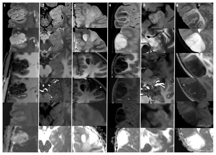
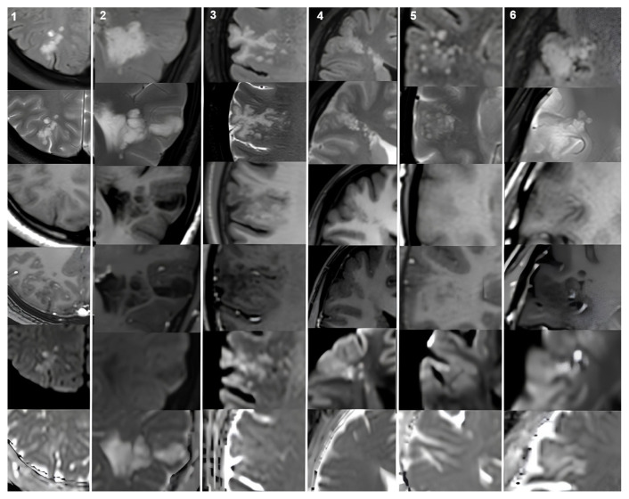
Results: There were 21 patients in the MVNT group and 20 patients in the DNET group. Headache was the most common symptom in patients with MVNTs (61.9%), whereas seizures were more prevalent in those with DNETs (70%). The most frequent locations for the MVNTs were the frontal and parietal lobes (66.6%), while DNETs were most commonly located in the temporal lobe (60%). All the MVNTs were hyperintense in both fluid-attenuated inversion recovery (FLAIR) and T2-weighted imaging (T2WI). All the DNETs were hyperintense on T2WI. However, on FLAIR, seven (35%) of the DNET lesions were hyperintense, while the remaining 13 lesions showed mixed signal intensity forming a bubbly appearance. Moreover, 20 of 21 (95.23%) MVNTs were hyperintense on diffusion-weighted imaging (DWI) (b800), with no apparent diffusion coefficient hypointensity in the lesions. None of the DNETs showed hyperintensity on DWI.
Conclusion: MRI findings, particularly those observed on FLAIR and DWI, may be helpful for distinguishing between MVNTs and DNETs, especially in cases where the differential diagnosis is challenging.
期刊介绍:
Turkish Journal of Medical sciences is a peer-reviewed comprehensive resource that provides critical up-to-date information on the broad spectrum of general medical sciences. The Journal intended to publish original medical scientific papers regarding the priority based on the prominence, significance, and timeliness of the findings. However since the audience of the Journal is not limited to any subspeciality in a wide variety of medical disciplines, the papers focusing on the technical details of a given medical subspeciality may not be evaluated for publication.
 求助内容:
求助内容: 应助结果提醒方式:


