Shijing Ma, Yingying Zhu, Changhong Pu, Jin Li, Bin Zhong
{"title":"计算机断层放射组学与肝细胞癌分化的临床参数相结合:一项机器学习研究。","authors":"Shijing Ma, Yingying Zhu, Changhong Pu, Jin Li, Bin Zhong","doi":"10.5114/pjr/200631","DOIUrl":null,"url":null,"abstract":"<p><strong>Purpose: </strong>To evaluate the performance of a combined clinical-radiomics model using multiple machine learning approaches for predicting pathological differentiation in hepatocellular carcinoma (HCC).</p><p><strong>Material and methods: </strong>A total of 196 patients with pathologically confirmed HCC, who underwent preoperative computed tomography (CT) were retrospectively enrolled (training: <i>n</i> = 156; validation: <i>n</i> = 40). The modelling process included the folowing: (1) clinical model construction through logistic regression analysis of risk factors; (2) radiomics model development by comparing 6 machine learning classifiers; and (3) integration of optimal clinical and radiomic features into a combined model. Model performance was assessed using the area under the curve (AUC), calibration curves, and decision curve analysis (DCA). A nomogram was constructed for clinical implementation.</p><p><strong>Results: </strong>Two clinical risk factors (BMI and CA153) were identified as independent predictors of differentiated HCC. The clinical model showed moderate performance (AUC: training = 0.705, validation = 0.658). The radiomics model demonstrated improved prediction capability (AUC: training = 0.840, validation = 0.716). The combined model achieved the best performance in differentiating HCC pathological grades (AUC: training = 0.878, validation = 0.747).</p><p><strong>Conclusions: </strong>The integration of CT radiomics features with clinical parameters through machine learning provides a promising non-invasive approach for predicting HCC pathological differentiation. This combined model could serve as a valuable tool for preoperative treatment planning.</p>","PeriodicalId":94174,"journal":{"name":"Polish journal of radiology","volume":"90 ","pages":"e140-e150"},"PeriodicalIF":0.0000,"publicationDate":"2025-03-24","publicationTypes":"Journal Article","fieldsOfStudy":null,"isOpenAccess":false,"openAccessPdf":"https://www.ncbi.nlm.nih.gov/pmc/articles/PMC12049157/pdf/","citationCount":"0","resultStr":"{\"title\":\"Computed tomography radiomics combined with clinical parameters for hepatocellular carcinoma differentiation: a machine learning investigation.\",\"authors\":\"Shijing Ma, Yingying Zhu, Changhong Pu, Jin Li, Bin Zhong\",\"doi\":\"10.5114/pjr/200631\",\"DOIUrl\":null,\"url\":null,\"abstract\":\"<p><strong>Purpose: </strong>To evaluate the performance of a combined clinical-radiomics model using multiple machine learning approaches for predicting pathological differentiation in hepatocellular carcinoma (HCC).</p><p><strong>Material and methods: </strong>A total of 196 patients with pathologically confirmed HCC, who underwent preoperative computed tomography (CT) were retrospectively enrolled (training: <i>n</i> = 156; validation: <i>n</i> = 40). The modelling process included the folowing: (1) clinical model construction through logistic regression analysis of risk factors; (2) radiomics model development by comparing 6 machine learning classifiers; and (3) integration of optimal clinical and radiomic features into a combined model. Model performance was assessed using the area under the curve (AUC), calibration curves, and decision curve analysis (DCA). A nomogram was constructed for clinical implementation.</p><p><strong>Results: </strong>Two clinical risk factors (BMI and CA153) were identified as independent predictors of differentiated HCC. The clinical model showed moderate performance (AUC: training = 0.705, validation = 0.658). The radiomics model demonstrated improved prediction capability (AUC: training = 0.840, validation = 0.716). The combined model achieved the best performance in differentiating HCC pathological grades (AUC: training = 0.878, validation = 0.747).</p><p><strong>Conclusions: </strong>The integration of CT radiomics features with clinical parameters through machine learning provides a promising non-invasive approach for predicting HCC pathological differentiation. This combined model could serve as a valuable tool for preoperative treatment planning.</p>\",\"PeriodicalId\":94174,\"journal\":{\"name\":\"Polish journal of radiology\",\"volume\":\"90 \",\"pages\":\"e140-e150\"},\"PeriodicalIF\":0.0000,\"publicationDate\":\"2025-03-24\",\"publicationTypes\":\"Journal Article\",\"fieldsOfStudy\":null,\"isOpenAccess\":false,\"openAccessPdf\":\"https://www.ncbi.nlm.nih.gov/pmc/articles/PMC12049157/pdf/\",\"citationCount\":\"0\",\"resultStr\":null,\"platform\":\"Semanticscholar\",\"paperid\":null,\"PeriodicalName\":\"Polish journal of radiology\",\"FirstCategoryId\":\"1085\",\"ListUrlMain\":\"https://doi.org/10.5114/pjr/200631\",\"RegionNum\":0,\"RegionCategory\":null,\"ArticlePicture\":[],\"TitleCN\":null,\"AbstractTextCN\":null,\"PMCID\":null,\"EPubDate\":\"2025/1/1 0:00:00\",\"PubModel\":\"eCollection\",\"JCR\":\"\",\"JCRName\":\"\",\"Score\":null,\"Total\":0}","platform":"Semanticscholar","paperid":null,"PeriodicalName":"Polish journal of radiology","FirstCategoryId":"1085","ListUrlMain":"https://doi.org/10.5114/pjr/200631","RegionNum":0,"RegionCategory":null,"ArticlePicture":[],"TitleCN":null,"AbstractTextCN":null,"PMCID":null,"EPubDate":"2025/1/1 0:00:00","PubModel":"eCollection","JCR":"","JCRName":"","Score":null,"Total":0}
引用次数: 0
摘要
目的:评估使用多种机器学习方法预测肝细胞癌(HCC)病理分化的临床-放射组学联合模型的性能。材料和方法:回顾性纳入196例经病理证实的HCC患者,术前行CT检查(training: n = 156;验证:n = 40)。建模过程包括:(1)通过危险因素的logistic回归分析构建临床模型;(2)通过比较6种机器学习分类器建立放射组学模型;(3)将最佳临床和放射学特征整合到一个组合模型中。使用曲线下面积(AUC)、校准曲线和决策曲线分析(DCA)评估模型性能。构建了临床应用的nomogram。结果:两个临床危险因素(BMI和CA153)被确定为分化型HCC的独立预测因素。临床模型表现中等(AUC: training = 0.705, validation = 0.658)。放射组学模型具有较好的预测能力(AUC: training = 0.840, validation = 0.716)。联合模型对HCC病理分级的鉴别效果最佳(AUC: training = 0.878, validation = 0.747)。结论:通过机器学习将CT放射组学特征与临床参数相结合,为HCC病理分化预测提供了一种有前景的无创方法。该组合模型可作为术前治疗计划的重要工具。
Computed tomography radiomics combined with clinical parameters for hepatocellular carcinoma differentiation: a machine learning investigation.
Purpose: To evaluate the performance of a combined clinical-radiomics model using multiple machine learning approaches for predicting pathological differentiation in hepatocellular carcinoma (HCC).
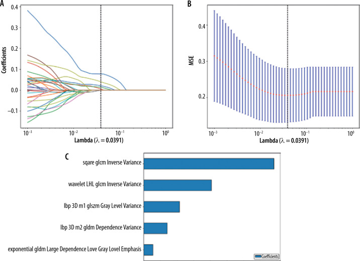
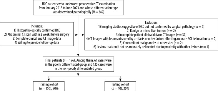
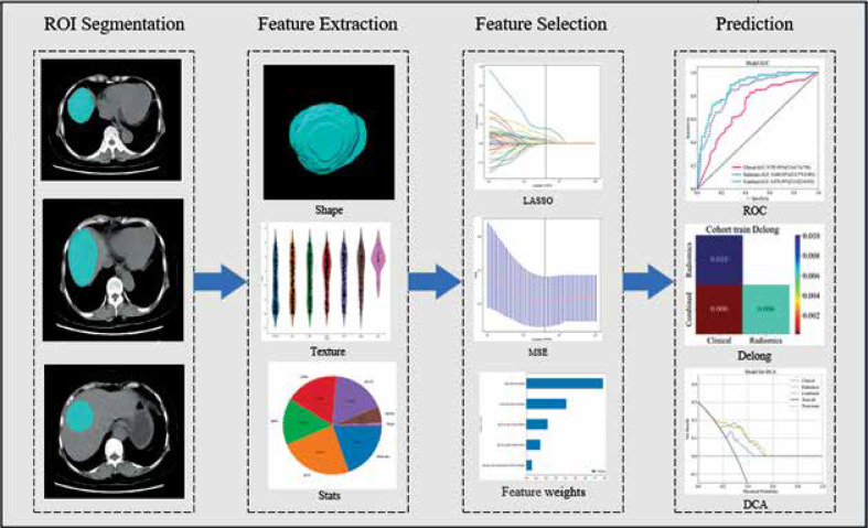
Material and methods: A total of 196 patients with pathologically confirmed HCC, who underwent preoperative computed tomography (CT) were retrospectively enrolled (training: n = 156; validation: n = 40). The modelling process included the folowing: (1) clinical model construction through logistic regression analysis of risk factors; (2) radiomics model development by comparing 6 machine learning classifiers; and (3) integration of optimal clinical and radiomic features into a combined model. Model performance was assessed using the area under the curve (AUC), calibration curves, and decision curve analysis (DCA). A nomogram was constructed for clinical implementation.
Results: Two clinical risk factors (BMI and CA153) were identified as independent predictors of differentiated HCC. The clinical model showed moderate performance (AUC: training = 0.705, validation = 0.658). The radiomics model demonstrated improved prediction capability (AUC: training = 0.840, validation = 0.716). The combined model achieved the best performance in differentiating HCC pathological grades (AUC: training = 0.878, validation = 0.747).
Conclusions: The integration of CT radiomics features with clinical parameters through machine learning provides a promising non-invasive approach for predicting HCC pathological differentiation. This combined model could serve as a valuable tool for preoperative treatment planning.

 求助内容:
求助内容: 应助结果提醒方式:


