{"title":"基于超声的多种腹部脂肪指标预测颈动脉粥样硬化。","authors":"Mohit Jain, Subhasish Panda, Shruti Chandak, Ankur Malhotra, Subhashree Dash, Umme Afifa","doi":"10.15557/JoU.2025.0006","DOIUrl":null,"url":null,"abstract":"<p><strong>Aim: </strong>Abdominal obesity is recognized as the best predictor of cardiovascular disease risk. While body mass index has traditionally been used to measure obesity, recent evidence suggests that visceral adipose tissue may be a better indicator of cardiovascular disease risk. Various surrogate imaging markers of visceral adipose tissue have recently been described, such as posterior right perinephric fat thickness, preperitoneal fat thickness, and the abdominal wall fat index. This study aimed to examine the link between atherosclerosis through measurement of carotid intima-media thickness and markers of abdominal obesity using ultrasonography.</p><p><strong>Material and methods: </strong>This was a hospital-based prospective observational study. Patients with a body mass index of 25-40 were included as cases and those with a body mass index 18.0-24.9 as controls. Posterior right perinephric fat thickness, preperitoneal fat thickness, and abdominal wall fat index were measured and compared with carotid intima-media thickness in cases.</p><p><strong>Results: </strong>A total of 100 cases and 100 age- and sex-matched controls were included. Body mass index did not show any statistically significant correlations with carotid intima-media thickness in this study. Among the visceral adiposity markers, posterior right perinephric fat thickness was the most sensitive and specific predictor of carotid intima-media thickness, while the abdominal wall fat index was the least sensitive and specific.</p><p><strong>Conclusions: </strong>Ultrasonographic markers of visceral adipose tissue, especially posterior right perinephric fat thickness and preperitoneal fat thickness, demonstrate a stronger association with carotid atherosclerosis than body mass index, making them useful predictors, particularly in individuals with high body mass index. These markers can be measured during routine abdominal ultrasounds to screen for atherosclerosis risk in patients with abdominal obesity.</p>","PeriodicalId":45612,"journal":{"name":"Journal of Ultrasonography","volume":"25 100","pages":"20250006"},"PeriodicalIF":1.5000,"publicationDate":"2025-03-26","publicationTypes":"Journal Article","fieldsOfStudy":null,"isOpenAccess":false,"openAccessPdf":"https://www.ncbi.nlm.nih.gov/pmc/articles/PMC11990943/pdf/","citationCount":"0","resultStr":"{\"title\":\"Ultrasonography-based prediction of carotid artery atherosclerosis using multiple abdominal fat indices.\",\"authors\":\"Mohit Jain, Subhasish Panda, Shruti Chandak, Ankur Malhotra, Subhashree Dash, Umme Afifa\",\"doi\":\"10.15557/JoU.2025.0006\",\"DOIUrl\":null,\"url\":null,\"abstract\":\"<p><strong>Aim: </strong>Abdominal obesity is recognized as the best predictor of cardiovascular disease risk. While body mass index has traditionally been used to measure obesity, recent evidence suggests that visceral adipose tissue may be a better indicator of cardiovascular disease risk. Various surrogate imaging markers of visceral adipose tissue have recently been described, such as posterior right perinephric fat thickness, preperitoneal fat thickness, and the abdominal wall fat index. This study aimed to examine the link between atherosclerosis through measurement of carotid intima-media thickness and markers of abdominal obesity using ultrasonography.</p><p><strong>Material and methods: </strong>This was a hospital-based prospective observational study. Patients with a body mass index of 25-40 were included as cases and those with a body mass index 18.0-24.9 as controls. Posterior right perinephric fat thickness, preperitoneal fat thickness, and abdominal wall fat index were measured and compared with carotid intima-media thickness in cases.</p><p><strong>Results: </strong>A total of 100 cases and 100 age- and sex-matched controls were included. Body mass index did not show any statistically significant correlations with carotid intima-media thickness in this study. Among the visceral adiposity markers, posterior right perinephric fat thickness was the most sensitive and specific predictor of carotid intima-media thickness, while the abdominal wall fat index was the least sensitive and specific.</p><p><strong>Conclusions: </strong>Ultrasonographic markers of visceral adipose tissue, especially posterior right perinephric fat thickness and preperitoneal fat thickness, demonstrate a stronger association with carotid atherosclerosis than body mass index, making them useful predictors, particularly in individuals with high body mass index. These markers can be measured during routine abdominal ultrasounds to screen for atherosclerosis risk in patients with abdominal obesity.</p>\",\"PeriodicalId\":45612,\"journal\":{\"name\":\"Journal of Ultrasonography\",\"volume\":\"25 100\",\"pages\":\"20250006\"},\"PeriodicalIF\":1.5000,\"publicationDate\":\"2025-03-26\",\"publicationTypes\":\"Journal Article\",\"fieldsOfStudy\":null,\"isOpenAccess\":false,\"openAccessPdf\":\"https://www.ncbi.nlm.nih.gov/pmc/articles/PMC11990943/pdf/\",\"citationCount\":\"0\",\"resultStr\":null,\"platform\":\"Semanticscholar\",\"paperid\":null,\"PeriodicalName\":\"Journal of Ultrasonography\",\"FirstCategoryId\":\"1085\",\"ListUrlMain\":\"https://doi.org/10.15557/JoU.2025.0006\",\"RegionNum\":0,\"RegionCategory\":null,\"ArticlePicture\":[],\"TitleCN\":null,\"AbstractTextCN\":null,\"PMCID\":null,\"EPubDate\":\"2025/1/1 0:00:00\",\"PubModel\":\"eCollection\",\"JCR\":\"Q3\",\"JCRName\":\"RADIOLOGY, NUCLEAR MEDICINE & MEDICAL IMAGING\",\"Score\":null,\"Total\":0}","platform":"Semanticscholar","paperid":null,"PeriodicalName":"Journal of Ultrasonography","FirstCategoryId":"1085","ListUrlMain":"https://doi.org/10.15557/JoU.2025.0006","RegionNum":0,"RegionCategory":null,"ArticlePicture":[],"TitleCN":null,"AbstractTextCN":null,"PMCID":null,"EPubDate":"2025/1/1 0:00:00","PubModel":"eCollection","JCR":"Q3","JCRName":"RADIOLOGY, NUCLEAR MEDICINE & MEDICAL IMAGING","Score":null,"Total":0}
Ultrasonography-based prediction of carotid artery atherosclerosis using multiple abdominal fat indices.
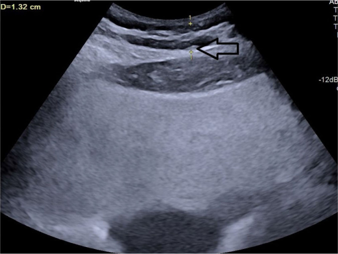
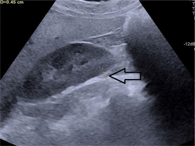
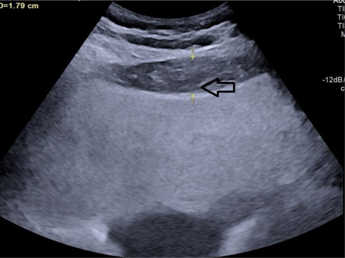
Aim: Abdominal obesity is recognized as the best predictor of cardiovascular disease risk. While body mass index has traditionally been used to measure obesity, recent evidence suggests that visceral adipose tissue may be a better indicator of cardiovascular disease risk. Various surrogate imaging markers of visceral adipose tissue have recently been described, such as posterior right perinephric fat thickness, preperitoneal fat thickness, and the abdominal wall fat index. This study aimed to examine the link between atherosclerosis through measurement of carotid intima-media thickness and markers of abdominal obesity using ultrasonography.
Material and methods: This was a hospital-based prospective observational study. Patients with a body mass index of 25-40 were included as cases and those with a body mass index 18.0-24.9 as controls. Posterior right perinephric fat thickness, preperitoneal fat thickness, and abdominal wall fat index were measured and compared with carotid intima-media thickness in cases.
Results: A total of 100 cases and 100 age- and sex-matched controls were included. Body mass index did not show any statistically significant correlations with carotid intima-media thickness in this study. Among the visceral adiposity markers, posterior right perinephric fat thickness was the most sensitive and specific predictor of carotid intima-media thickness, while the abdominal wall fat index was the least sensitive and specific.
Conclusions: Ultrasonographic markers of visceral adipose tissue, especially posterior right perinephric fat thickness and preperitoneal fat thickness, demonstrate a stronger association with carotid atherosclerosis than body mass index, making them useful predictors, particularly in individuals with high body mass index. These markers can be measured during routine abdominal ultrasounds to screen for atherosclerosis risk in patients with abdominal obesity.

 求助内容:
求助内容: 应助结果提醒方式:


