Francesca Castagnoli, Mihaela Rata, Joshua Shur, Georgina Hopkinson, Alison Macdonald, David Stockton, Marcel Dominik Nickel, Stephan Kannengiesser, Christina Messiou, Dow-Mu Koh, Jessica Mary Winfield
{"title":"人工智能增强重建提供了更好的图像质量,并使对比增强肝脏MRI的屏气时间更短。","authors":"Francesca Castagnoli, Mihaela Rata, Joshua Shur, Georgina Hopkinson, Alison Macdonald, David Stockton, Marcel Dominik Nickel, Stephan Kannengiesser, Christina Messiou, Dow-Mu Koh, Jessica Mary Winfield","doi":"10.1186/s41747-025-00582-1","DOIUrl":null,"url":null,"abstract":"<p><strong>Background: </strong>To compare liver image quality and lesion detection using an AI-augmented T1-weighted sequence on hepatobiliary-phase gadoxetate-enhanced magnetic resonance imaging (MRI).</p><p><strong>Methods: </strong>Fifty patients undergoing gadoxetate-enhanced MRI were recruited. Two T1-weighted Dixon sequences were utilized: a 17-s breath-hold acquisition and an accelerated 12-s breath-hold acquisition (reduced phase resolution), both reconstructed using neural network (NN) and iterative denoising (ID), NN-alone, ID-alone, and the standard method. Contrast-to-noise ratio (CNR) was assessed quantitatively for all series (ANOVA). Two blinded radiologists independently analyzed three image sets: 17-s acquisition reconstructed with NN and ID (17-s NN + ID), 12-s acquisition reconstructed with NN and ID (12-s NN + ID), and 17-s acquisition with standard reconstruction (17-s standard). Overall image quality, qualitative CNR, lesion edge sharpness, vessel edge sharpness, and respiratory motion artifacts were scored (4-point Likert scale) and compared (Friedman test). Lesion detection was compared between 12-s NN + ID and 17-s standard reconstructions (Wilcoxon signed-rank test).</p><p><strong>Results: </strong>Quantitative liver-to-portal vein CNR was significantly higher for 17-s NN + ID than 17-s standard or 17-s NN-alone images (p = 0.001). Scores for overall image quality, qualitative CNR, vessel edge sharpness, and lesion edge sharpness were significantly higher for 17-s NN + ID and 12-s NN + ID than standard reconstruction (p < 0.001); there was no significant difference between 17-s and 12-s NN + ID. There was no significant difference in respiratory motion artifacts and number of lesions or diameter of the smallest detected lesion using 12-s NN + ID or 17-s standard reconstruction.</p><p><strong>Conclusion: </strong>AI-augmented reconstructions can improve image quality while reducing breath-hold duration in T1-weighted hepatobiliary-phase gadoxetate-enhanced MRI, without compromising lesion detection.</p><p><strong>Relevance statement: </strong>AI-augmented reconstruction of T1-weighted MRI improves image quality and lesion detection in hepatobiliary phase liver imaging, reducing breath-hold duration without compromising clinical lesion detection.</p><p><strong>Key points: </strong>Liver-to-portal vein CNR was significantly higher for 17-s NN + ID. AI-augmented reconstructions scored higher for image quality, contrast-to-noise, vessel-edge, and lesion-edge sharpness. No significant difference in lesion detection between 12-s NN + ID and 17-s standard reconstructions.</p>","PeriodicalId":36926,"journal":{"name":"European Radiology Experimental","volume":"9 1","pages":"46"},"PeriodicalIF":3.6000,"publicationDate":"2025-05-01","publicationTypes":"Journal Article","fieldsOfStudy":null,"isOpenAccess":false,"openAccessPdf":"https://www.ncbi.nlm.nih.gov/pmc/articles/PMC12045906/pdf/","citationCount":"0","resultStr":"{\"title\":\"AI-augmented reconstruction provides improved image quality and enables shorter breath-holds in contrast-enhanced liver MRI.\",\"authors\":\"Francesca Castagnoli, Mihaela Rata, Joshua Shur, Georgina Hopkinson, Alison Macdonald, David Stockton, Marcel Dominik Nickel, Stephan Kannengiesser, Christina Messiou, Dow-Mu Koh, Jessica Mary Winfield\",\"doi\":\"10.1186/s41747-025-00582-1\",\"DOIUrl\":null,\"url\":null,\"abstract\":\"<p><strong>Background: </strong>To compare liver image quality and lesion detection using an AI-augmented T1-weighted sequence on hepatobiliary-phase gadoxetate-enhanced magnetic resonance imaging (MRI).</p><p><strong>Methods: </strong>Fifty patients undergoing gadoxetate-enhanced MRI were recruited. Two T1-weighted Dixon sequences were utilized: a 17-s breath-hold acquisition and an accelerated 12-s breath-hold acquisition (reduced phase resolution), both reconstructed using neural network (NN) and iterative denoising (ID), NN-alone, ID-alone, and the standard method. Contrast-to-noise ratio (CNR) was assessed quantitatively for all series (ANOVA). Two blinded radiologists independently analyzed three image sets: 17-s acquisition reconstructed with NN and ID (17-s NN + ID), 12-s acquisition reconstructed with NN and ID (12-s NN + ID), and 17-s acquisition with standard reconstruction (17-s standard). Overall image quality, qualitative CNR, lesion edge sharpness, vessel edge sharpness, and respiratory motion artifacts were scored (4-point Likert scale) and compared (Friedman test). Lesion detection was compared between 12-s NN + ID and 17-s standard reconstructions (Wilcoxon signed-rank test).</p><p><strong>Results: </strong>Quantitative liver-to-portal vein CNR was significantly higher for 17-s NN + ID than 17-s standard or 17-s NN-alone images (p = 0.001). Scores for overall image quality, qualitative CNR, vessel edge sharpness, and lesion edge sharpness were significantly higher for 17-s NN + ID and 12-s NN + ID than standard reconstruction (p < 0.001); there was no significant difference between 17-s and 12-s NN + ID. There was no significant difference in respiratory motion artifacts and number of lesions or diameter of the smallest detected lesion using 12-s NN + ID or 17-s standard reconstruction.</p><p><strong>Conclusion: </strong>AI-augmented reconstructions can improve image quality while reducing breath-hold duration in T1-weighted hepatobiliary-phase gadoxetate-enhanced MRI, without compromising lesion detection.</p><p><strong>Relevance statement: </strong>AI-augmented reconstruction of T1-weighted MRI improves image quality and lesion detection in hepatobiliary phase liver imaging, reducing breath-hold duration without compromising clinical lesion detection.</p><p><strong>Key points: </strong>Liver-to-portal vein CNR was significantly higher for 17-s NN + ID. AI-augmented reconstructions scored higher for image quality, contrast-to-noise, vessel-edge, and lesion-edge sharpness. No significant difference in lesion detection between 12-s NN + ID and 17-s standard reconstructions.</p>\",\"PeriodicalId\":36926,\"journal\":{\"name\":\"European Radiology Experimental\",\"volume\":\"9 1\",\"pages\":\"46\"},\"PeriodicalIF\":3.6000,\"publicationDate\":\"2025-05-01\",\"publicationTypes\":\"Journal Article\",\"fieldsOfStudy\":null,\"isOpenAccess\":false,\"openAccessPdf\":\"https://www.ncbi.nlm.nih.gov/pmc/articles/PMC12045906/pdf/\",\"citationCount\":\"0\",\"resultStr\":null,\"platform\":\"Semanticscholar\",\"paperid\":null,\"PeriodicalName\":\"European Radiology Experimental\",\"FirstCategoryId\":\"1085\",\"ListUrlMain\":\"https://doi.org/10.1186/s41747-025-00582-1\",\"RegionNum\":0,\"RegionCategory\":null,\"ArticlePicture\":[],\"TitleCN\":null,\"AbstractTextCN\":null,\"PMCID\":null,\"EPubDate\":\"\",\"PubModel\":\"\",\"JCR\":\"Q1\",\"JCRName\":\"RADIOLOGY, NUCLEAR MEDICINE & MEDICAL IMAGING\",\"Score\":null,\"Total\":0}","platform":"Semanticscholar","paperid":null,"PeriodicalName":"European Radiology Experimental","FirstCategoryId":"1085","ListUrlMain":"https://doi.org/10.1186/s41747-025-00582-1","RegionNum":0,"RegionCategory":null,"ArticlePicture":[],"TitleCN":null,"AbstractTextCN":null,"PMCID":null,"EPubDate":"","PubModel":"","JCR":"Q1","JCRName":"RADIOLOGY, NUCLEAR MEDICINE & MEDICAL IMAGING","Score":null,"Total":0}
引用次数: 0
摘要
背景:比较人工智能增强肝胆道期加多赛特增强磁共振成像(MRI)的肝脏图像质量和病变检测。方法:招募50例接受加多赛特增强MRI检查的患者。使用两个t1加权Dixon序列:17秒屏气采集和加速12秒屏气采集(降低相位分辨率),均使用神经网络(NN)和迭代去噪(ID),单独使用NN,单独使用ID和标准方法重建。定量评估所有系列的噪比(CNR) (ANOVA)。两名盲法放射科医生独立分析了三组图像:用神经网络和ID重建的17-s采集(17-s NN + ID)、用神经网络和ID重建的12-s采集(12-s NN + ID)和用标准重建的17-s采集(17-s标准)。对整体图像质量、定性CNR、病变边缘清晰度、血管边缘清晰度和呼吸运动伪影进行评分(4点李克特量表)并进行比较(弗里德曼检验)。比较12 s NN + ID和17 s标准重建的病变检测(Wilcoxon sign -rank检验)。结果:17-s NN + ID的定量肝到门静脉CNR明显高于17-s标准图像或17-s NN单独图像(p = 0.001)。17-s NN + ID和12-s NN + ID的整体图像质量、定性CNR、血管边缘清晰度和病变边缘清晰度评分明显高于标准重建(p)。结论:人工智能增强重建可以改善图像质量,同时减少t1加权肝胆道期加多酸酯增强MRI的屏气时间,而不影响病变检测。相关声明:人工智能增强的t1加权MRI重建改善了肝胆期肝脏成像的图像质量和病变检测,减少了屏气时间,但不影响临床病变检测。重点:17 s NN + ID肝至门静脉CNR明显增高。人工智能增强重建在图像质量、噪声对比度、血管边缘和病变边缘清晰度方面得分更高。12-s NN + ID与17-s标准重建的病变检出率无显著差异。
AI-augmented reconstruction provides improved image quality and enables shorter breath-holds in contrast-enhanced liver MRI.
Background: To compare liver image quality and lesion detection using an AI-augmented T1-weighted sequence on hepatobiliary-phase gadoxetate-enhanced magnetic resonance imaging (MRI).
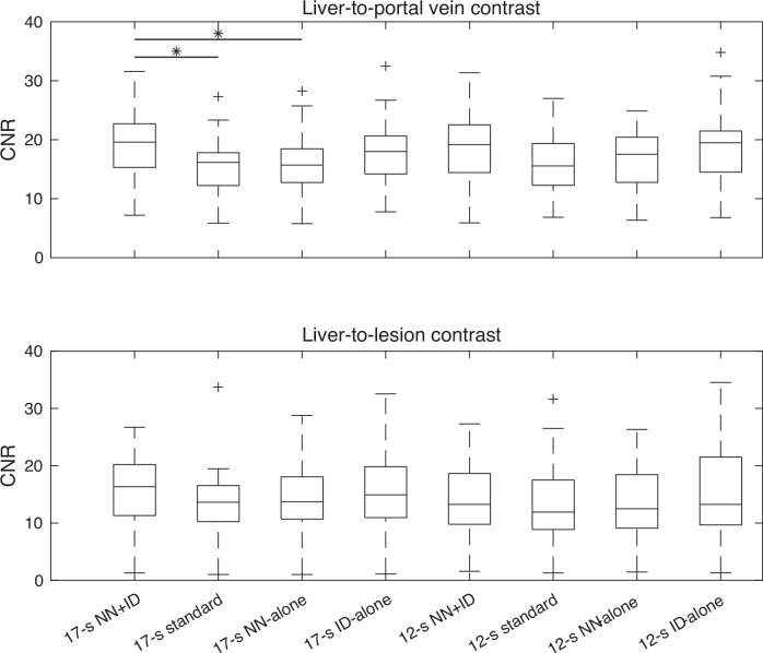
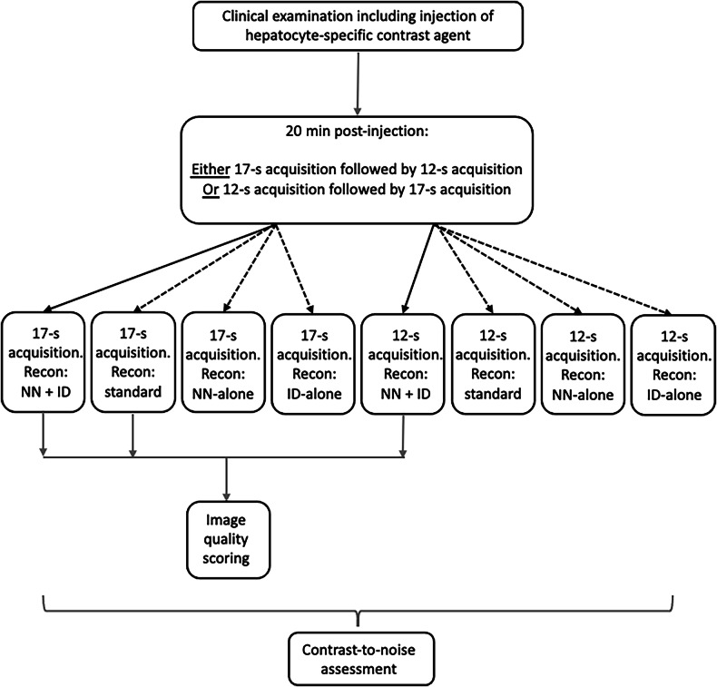
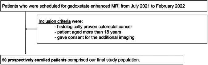
Methods: Fifty patients undergoing gadoxetate-enhanced MRI were recruited. Two T1-weighted Dixon sequences were utilized: a 17-s breath-hold acquisition and an accelerated 12-s breath-hold acquisition (reduced phase resolution), both reconstructed using neural network (NN) and iterative denoising (ID), NN-alone, ID-alone, and the standard method. Contrast-to-noise ratio (CNR) was assessed quantitatively for all series (ANOVA). Two blinded radiologists independently analyzed three image sets: 17-s acquisition reconstructed with NN and ID (17-s NN + ID), 12-s acquisition reconstructed with NN and ID (12-s NN + ID), and 17-s acquisition with standard reconstruction (17-s standard). Overall image quality, qualitative CNR, lesion edge sharpness, vessel edge sharpness, and respiratory motion artifacts were scored (4-point Likert scale) and compared (Friedman test). Lesion detection was compared between 12-s NN + ID and 17-s standard reconstructions (Wilcoxon signed-rank test).
Results: Quantitative liver-to-portal vein CNR was significantly higher for 17-s NN + ID than 17-s standard or 17-s NN-alone images (p = 0.001). Scores for overall image quality, qualitative CNR, vessel edge sharpness, and lesion edge sharpness were significantly higher for 17-s NN + ID and 12-s NN + ID than standard reconstruction (p < 0.001); there was no significant difference between 17-s and 12-s NN + ID. There was no significant difference in respiratory motion artifacts and number of lesions or diameter of the smallest detected lesion using 12-s NN + ID or 17-s standard reconstruction.
Conclusion: AI-augmented reconstructions can improve image quality while reducing breath-hold duration in T1-weighted hepatobiliary-phase gadoxetate-enhanced MRI, without compromising lesion detection.
Relevance statement: AI-augmented reconstruction of T1-weighted MRI improves image quality and lesion detection in hepatobiliary phase liver imaging, reducing breath-hold duration without compromising clinical lesion detection.
Key points: Liver-to-portal vein CNR was significantly higher for 17-s NN + ID. AI-augmented reconstructions scored higher for image quality, contrast-to-noise, vessel-edge, and lesion-edge sharpness. No significant difference in lesion detection between 12-s NN + ID and 17-s standard reconstructions.

 求助内容:
求助内容: 应助结果提醒方式:


