{"title":"不同葡萄膜条件下超宽视场荧光素血管造影中的蕨类图案。","authors":"Vipin Rana, Meenu Dangi, Pradeep Kumar, Ashish Markan, Jaya Kaushik, Ranjit Goenka, Anupama Rana, Atul Gupta, Amit Nandan Tripathi","doi":"10.22336/rjo.2025.16","DOIUrl":null,"url":null,"abstract":"<p><strong>Objective: </strong>To investigate the occurrence and characteristics of the \"Fern-like pattern\" (FLP) on ultra-wide field fluorescein angiography (UWFA) across a variety of uveitic conditions, challenging the traditional association of FLP primarily with Behçet's disease (BD).</p><p><strong>Methods: </strong>This observational study at a tertiary care centre in India analysed the UWFA images of 23 eyes from 12 patients diagnosed with BD, tubercular vasculitis, intermediate uveitis, Vogt‒Koyanagi‒Harada (VKH) syndrome, or exudative retinal detachment. Clinical and imaging data were reviewed to assess the presence and implications of FLP in these conditions.</p><p><strong>Results: </strong>The study included 12 patients: 8 males (66.67%) and four females (33.33%). The distribution was: 3 with BD, 4 with tubercular vasculitis, 3 with intermediate uveitis, 1 with VKH syndrome, and 1 with exudative retinal detachment. The average age was 29.58 ± 11.35 years. Anterior segment examination revealed significantly more hypopyon, cells, and posterior synechiae in BD patients. Posterior segment examination showed choroiditis exclusively in non-Behçet's patients and considerably increased vitritis in BD patients. Disc hyperfluorescence and neovascularization were substantially more common in BD patients.</p><p><strong>Discussion: </strong>Our study is the first to comprehensively assess the presence of FLP on UWFA across both BD and non-BD uveitic conditions, demonstrating its occurrence in TB vasculitis, VKH syndrome, and intermediate uveitis. While traditionally linked to BD, FLP signifies retinal capillary leakage and inflammation, necessitating careful differential diagnosis. We observed optic disc hyperfluorescence predominantly in BD, underscoring its role as a potential disease activity marker requiring aggressive immunosuppression. Notably, FLP was identified in exudative retinal detachment and VKH, suggesting shared inflammatory mechanisms across conditions. These findings broaden the clinical relevance of FLP in uveitis and highlight the need for further studies to explore its diagnostic and prognostic significance.</p><p><strong>Conclusion: </strong>The study confirms that FLP on UWFA is not exclusive to BD and appears in other uveitic conditions. Clinical signs such as hypopyon and disc hyperfluorescence indicate BD, whereas choroiditis is more common in non-Behçet's uveitis. These findings highlight the importance of precise clinical and imaging assessments to distinguish between different uveitic conditions for accurate diagnosis and effective treatment.</p>","PeriodicalId":94355,"journal":{"name":"Romanian journal of ophthalmology","volume":"69 1","pages":"101-109"},"PeriodicalIF":0.0000,"publicationDate":"2025-01-01","publicationTypes":"Journal Article","fieldsOfStudy":null,"isOpenAccess":false,"openAccessPdf":"https://www.ncbi.nlm.nih.gov/pmc/articles/PMC12049652/pdf/","citationCount":"0","resultStr":"{\"title\":\"Fern-Like Pattern on Ultra-Widefield Fluorescein Angiography Across Various Uveitic Conditions.\",\"authors\":\"Vipin Rana, Meenu Dangi, Pradeep Kumar, Ashish Markan, Jaya Kaushik, Ranjit Goenka, Anupama Rana, Atul Gupta, Amit Nandan Tripathi\",\"doi\":\"10.22336/rjo.2025.16\",\"DOIUrl\":null,\"url\":null,\"abstract\":\"<p><strong>Objective: </strong>To investigate the occurrence and characteristics of the \\\"Fern-like pattern\\\" (FLP) on ultra-wide field fluorescein angiography (UWFA) across a variety of uveitic conditions, challenging the traditional association of FLP primarily with Behçet's disease (BD).</p><p><strong>Methods: </strong>This observational study at a tertiary care centre in India analysed the UWFA images of 23 eyes from 12 patients diagnosed with BD, tubercular vasculitis, intermediate uveitis, Vogt‒Koyanagi‒Harada (VKH) syndrome, or exudative retinal detachment. Clinical and imaging data were reviewed to assess the presence and implications of FLP in these conditions.</p><p><strong>Results: </strong>The study included 12 patients: 8 males (66.67%) and four females (33.33%). The distribution was: 3 with BD, 4 with tubercular vasculitis, 3 with intermediate uveitis, 1 with VKH syndrome, and 1 with exudative retinal detachment. The average age was 29.58 ± 11.35 years. Anterior segment examination revealed significantly more hypopyon, cells, and posterior synechiae in BD patients. Posterior segment examination showed choroiditis exclusively in non-Behçet's patients and considerably increased vitritis in BD patients. Disc hyperfluorescence and neovascularization were substantially more common in BD patients.</p><p><strong>Discussion: </strong>Our study is the first to comprehensively assess the presence of FLP on UWFA across both BD and non-BD uveitic conditions, demonstrating its occurrence in TB vasculitis, VKH syndrome, and intermediate uveitis. While traditionally linked to BD, FLP signifies retinal capillary leakage and inflammation, necessitating careful differential diagnosis. We observed optic disc hyperfluorescence predominantly in BD, underscoring its role as a potential disease activity marker requiring aggressive immunosuppression. Notably, FLP was identified in exudative retinal detachment and VKH, suggesting shared inflammatory mechanisms across conditions. These findings broaden the clinical relevance of FLP in uveitis and highlight the need for further studies to explore its diagnostic and prognostic significance.</p><p><strong>Conclusion: </strong>The study confirms that FLP on UWFA is not exclusive to BD and appears in other uveitic conditions. Clinical signs such as hypopyon and disc hyperfluorescence indicate BD, whereas choroiditis is more common in non-Behçet's uveitis. These findings highlight the importance of precise clinical and imaging assessments to distinguish between different uveitic conditions for accurate diagnosis and effective treatment.</p>\",\"PeriodicalId\":94355,\"journal\":{\"name\":\"Romanian journal of ophthalmology\",\"volume\":\"69 1\",\"pages\":\"101-109\"},\"PeriodicalIF\":0.0000,\"publicationDate\":\"2025-01-01\",\"publicationTypes\":\"Journal Article\",\"fieldsOfStudy\":null,\"isOpenAccess\":false,\"openAccessPdf\":\"https://www.ncbi.nlm.nih.gov/pmc/articles/PMC12049652/pdf/\",\"citationCount\":\"0\",\"resultStr\":null,\"platform\":\"Semanticscholar\",\"paperid\":null,\"PeriodicalName\":\"Romanian journal of ophthalmology\",\"FirstCategoryId\":\"1085\",\"ListUrlMain\":\"https://doi.org/10.22336/rjo.2025.16\",\"RegionNum\":0,\"RegionCategory\":null,\"ArticlePicture\":[],\"TitleCN\":null,\"AbstractTextCN\":null,\"PMCID\":null,\"EPubDate\":\"\",\"PubModel\":\"\",\"JCR\":\"\",\"JCRName\":\"\",\"Score\":null,\"Total\":0}","platform":"Semanticscholar","paperid":null,"PeriodicalName":"Romanian journal of ophthalmology","FirstCategoryId":"1085","ListUrlMain":"https://doi.org/10.22336/rjo.2025.16","RegionNum":0,"RegionCategory":null,"ArticlePicture":[],"TitleCN":null,"AbstractTextCN":null,"PMCID":null,"EPubDate":"","PubModel":"","JCR":"","JCRName":"","Score":null,"Total":0}
Fern-Like Pattern on Ultra-Widefield Fluorescein Angiography Across Various Uveitic Conditions.
Objective: To investigate the occurrence and characteristics of the "Fern-like pattern" (FLP) on ultra-wide field fluorescein angiography (UWFA) across a variety of uveitic conditions, challenging the traditional association of FLP primarily with Behçet's disease (BD).
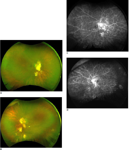
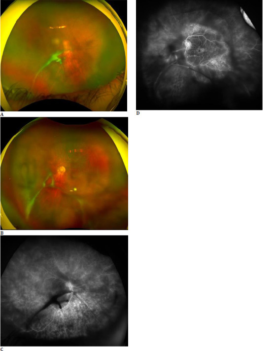
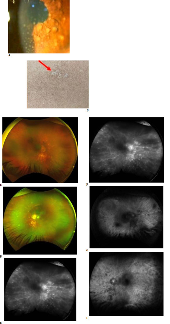
Methods: This observational study at a tertiary care centre in India analysed the UWFA images of 23 eyes from 12 patients diagnosed with BD, tubercular vasculitis, intermediate uveitis, Vogt‒Koyanagi‒Harada (VKH) syndrome, or exudative retinal detachment. Clinical and imaging data were reviewed to assess the presence and implications of FLP in these conditions.
Results: The study included 12 patients: 8 males (66.67%) and four females (33.33%). The distribution was: 3 with BD, 4 with tubercular vasculitis, 3 with intermediate uveitis, 1 with VKH syndrome, and 1 with exudative retinal detachment. The average age was 29.58 ± 11.35 years. Anterior segment examination revealed significantly more hypopyon, cells, and posterior synechiae in BD patients. Posterior segment examination showed choroiditis exclusively in non-Behçet's patients and considerably increased vitritis in BD patients. Disc hyperfluorescence and neovascularization were substantially more common in BD patients.
Discussion: Our study is the first to comprehensively assess the presence of FLP on UWFA across both BD and non-BD uveitic conditions, demonstrating its occurrence in TB vasculitis, VKH syndrome, and intermediate uveitis. While traditionally linked to BD, FLP signifies retinal capillary leakage and inflammation, necessitating careful differential diagnosis. We observed optic disc hyperfluorescence predominantly in BD, underscoring its role as a potential disease activity marker requiring aggressive immunosuppression. Notably, FLP was identified in exudative retinal detachment and VKH, suggesting shared inflammatory mechanisms across conditions. These findings broaden the clinical relevance of FLP in uveitis and highlight the need for further studies to explore its diagnostic and prognostic significance.
Conclusion: The study confirms that FLP on UWFA is not exclusive to BD and appears in other uveitic conditions. Clinical signs such as hypopyon and disc hyperfluorescence indicate BD, whereas choroiditis is more common in non-Behçet's uveitis. These findings highlight the importance of precise clinical and imaging assessments to distinguish between different uveitic conditions for accurate diagnosis and effective treatment.

 求助内容:
求助内容: 应助结果提醒方式:


