{"title":"FROP-1肽偶联的超顺磁性纳米颗粒作为乳腺癌靶向t1加权MR造影剂:体外研究","authors":"Melika Samari, Zahra Alamzadeh, Rasoul Irajirad, Abolfazl Sarikhani, Vahid Pirhajati Mahabadi, Habib Ghaznavi, Samideh Khoei","doi":"10.1186/s42490-025-00091-7","DOIUrl":null,"url":null,"abstract":"<p><strong>Background: </strong>The aim of this study was to produce ultrasmall superparamagnetic iron oxide (USPIO) nanoparticles (NPs) conjugated to the FROP-1 peptide for targeted magnetic resonance imaging (MRI) of breast cancer cell lines and to evaluate its application as a specific and targeted T1-weighted MR imaging contrast agent in vitro. Sodium citrate-stabilized Fe<sub>3</sub>O<sub>4</sub> NPs were conjugated with the FROP-1 peptide by 1-ethyl-3-(3-dimethylaminopropyl) carbide diamide hydrochloride (EDC) to form a novel Fe<sub>3</sub>O<sub>4</sub>@FROP-1 specific target contrast agent. The specificity and targeting of Fe<sub>3</sub>O<sub>4</sub>@FROP-1 to bind FROP-1 receptors were investigated in vitro by cellular uptake and cellular MR imaging.</p><p><strong>Results: </strong>In this study, the synthesis of water-soluble ultrasmall Fe<sub>3</sub>O<sub>4</sub> NPs was performed by the co-precipitation method. XRD, TEM, and VSM analyses showed the formation of the Fe<sub>3</sub>O<sub>4</sub> NPs with an average size of about 3.78 ± 0.2 nm. FT-IR spectroscopy approved the conjugation of the FROP-1 peptide with the Fe<sub>3</sub>O<sub>4</sub> NPs. The synthesized Fe<sub>3</sub>O<sub>4</sub>@FROP-1 NPs showed good biocompatibility, and the high r1 relaxivity and r2/r1, respectively, were 2.608 mM<sup>- 1</sup>S<sup>- 1</sup> and 1.18. The biocompatibility of the Fe<sub>3</sub>O<sub>4</sub> and Fe<sub>3</sub>O<sub>4</sub>@FROP-1 NPs on the MCF-7, SKBR-3, MDA-MB-231, and MCF-10 cell lines was determined using cytotoxicity analysis. The specific targeting effect on the cells was verified by in vitro cellular uptake and cell MR imaging.</p><p><strong>Conclusion: </strong>It was found that the contrast intensity of the Fe<sub>3</sub>O<sub>4</sub>@FROP-1 nanoprobe increases as Fe concentration increases. Cellular uptake of the Fe<sub>3</sub>O<sub>4</sub> and Fe<sub>3</sub>O<sub>4</sub>@FROP-1 NPs was quantified using ICP-MS. The synthesized NPs had better imaging performance than Dotarem (gadoterate meglumine). The findings showed that Fe<sub>3</sub>O<sub>4</sub>@FROP-1 NPs have potential utility as a specific and targeted T1-weighted contrast agent in breast cancer MR imaging.</p>","PeriodicalId":72425,"journal":{"name":"BMC biomedical engineering","volume":"7 1","pages":"5"},"PeriodicalIF":0.0000,"publicationDate":"2025-05-01","publicationTypes":"Journal Article","fieldsOfStudy":null,"isOpenAccess":false,"openAccessPdf":"https://www.ncbi.nlm.nih.gov/pmc/articles/PMC12044754/pdf/","citationCount":"0","resultStr":"{\"title\":\"FROP-1 peptide-conjugated ultrasmall superparamagnetic nanoparticles as a targeted T1-weighted MR contrast agent for breast cancer: in vitro study.\",\"authors\":\"Melika Samari, Zahra Alamzadeh, Rasoul Irajirad, Abolfazl Sarikhani, Vahid Pirhajati Mahabadi, Habib Ghaznavi, Samideh Khoei\",\"doi\":\"10.1186/s42490-025-00091-7\",\"DOIUrl\":null,\"url\":null,\"abstract\":\"<p><strong>Background: </strong>The aim of this study was to produce ultrasmall superparamagnetic iron oxide (USPIO) nanoparticles (NPs) conjugated to the FROP-1 peptide for targeted magnetic resonance imaging (MRI) of breast cancer cell lines and to evaluate its application as a specific and targeted T1-weighted MR imaging contrast agent in vitro. Sodium citrate-stabilized Fe<sub>3</sub>O<sub>4</sub> NPs were conjugated with the FROP-1 peptide by 1-ethyl-3-(3-dimethylaminopropyl) carbide diamide hydrochloride (EDC) to form a novel Fe<sub>3</sub>O<sub>4</sub>@FROP-1 specific target contrast agent. The specificity and targeting of Fe<sub>3</sub>O<sub>4</sub>@FROP-1 to bind FROP-1 receptors were investigated in vitro by cellular uptake and cellular MR imaging.</p><p><strong>Results: </strong>In this study, the synthesis of water-soluble ultrasmall Fe<sub>3</sub>O<sub>4</sub> NPs was performed by the co-precipitation method. XRD, TEM, and VSM analyses showed the formation of the Fe<sub>3</sub>O<sub>4</sub> NPs with an average size of about 3.78 ± 0.2 nm. FT-IR spectroscopy approved the conjugation of the FROP-1 peptide with the Fe<sub>3</sub>O<sub>4</sub> NPs. The synthesized Fe<sub>3</sub>O<sub>4</sub>@FROP-1 NPs showed good biocompatibility, and the high r1 relaxivity and r2/r1, respectively, were 2.608 mM<sup>- 1</sup>S<sup>- 1</sup> and 1.18. The biocompatibility of the Fe<sub>3</sub>O<sub>4</sub> and Fe<sub>3</sub>O<sub>4</sub>@FROP-1 NPs on the MCF-7, SKBR-3, MDA-MB-231, and MCF-10 cell lines was determined using cytotoxicity analysis. The specific targeting effect on the cells was verified by in vitro cellular uptake and cell MR imaging.</p><p><strong>Conclusion: </strong>It was found that the contrast intensity of the Fe<sub>3</sub>O<sub>4</sub>@FROP-1 nanoprobe increases as Fe concentration increases. Cellular uptake of the Fe<sub>3</sub>O<sub>4</sub> and Fe<sub>3</sub>O<sub>4</sub>@FROP-1 NPs was quantified using ICP-MS. The synthesized NPs had better imaging performance than Dotarem (gadoterate meglumine). The findings showed that Fe<sub>3</sub>O<sub>4</sub>@FROP-1 NPs have potential utility as a specific and targeted T1-weighted contrast agent in breast cancer MR imaging.</p>\",\"PeriodicalId\":72425,\"journal\":{\"name\":\"BMC biomedical engineering\",\"volume\":\"7 1\",\"pages\":\"5\"},\"PeriodicalIF\":0.0000,\"publicationDate\":\"2025-05-01\",\"publicationTypes\":\"Journal Article\",\"fieldsOfStudy\":null,\"isOpenAccess\":false,\"openAccessPdf\":\"https://www.ncbi.nlm.nih.gov/pmc/articles/PMC12044754/pdf/\",\"citationCount\":\"0\",\"resultStr\":null,\"platform\":\"Semanticscholar\",\"paperid\":null,\"PeriodicalName\":\"BMC biomedical engineering\",\"FirstCategoryId\":\"1085\",\"ListUrlMain\":\"https://doi.org/10.1186/s42490-025-00091-7\",\"RegionNum\":0,\"RegionCategory\":null,\"ArticlePicture\":[],\"TitleCN\":null,\"AbstractTextCN\":null,\"PMCID\":null,\"EPubDate\":\"\",\"PubModel\":\"\",\"JCR\":\"\",\"JCRName\":\"\",\"Score\":null,\"Total\":0}","platform":"Semanticscholar","paperid":null,"PeriodicalName":"BMC biomedical engineering","FirstCategoryId":"1085","ListUrlMain":"https://doi.org/10.1186/s42490-025-00091-7","RegionNum":0,"RegionCategory":null,"ArticlePicture":[],"TitleCN":null,"AbstractTextCN":null,"PMCID":null,"EPubDate":"","PubModel":"","JCR":"","JCRName":"","Score":null,"Total":0}
FROP-1 peptide-conjugated ultrasmall superparamagnetic nanoparticles as a targeted T1-weighted MR contrast agent for breast cancer: in vitro study.
Background: The aim of this study was to produce ultrasmall superparamagnetic iron oxide (USPIO) nanoparticles (NPs) conjugated to the FROP-1 peptide for targeted magnetic resonance imaging (MRI) of breast cancer cell lines and to evaluate its application as a specific and targeted T1-weighted MR imaging contrast agent in vitro. Sodium citrate-stabilized Fe3O4 NPs were conjugated with the FROP-1 peptide by 1-ethyl-3-(3-dimethylaminopropyl) carbide diamide hydrochloride (EDC) to form a novel Fe3O4@FROP-1 specific target contrast agent. The specificity and targeting of Fe3O4@FROP-1 to bind FROP-1 receptors were investigated in vitro by cellular uptake and cellular MR imaging.
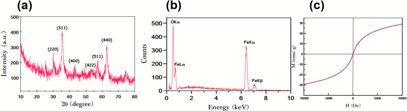
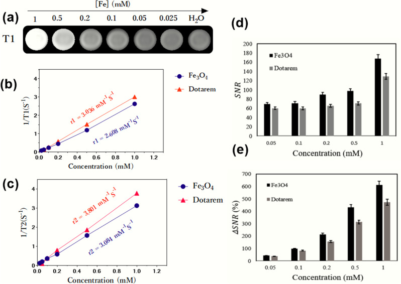
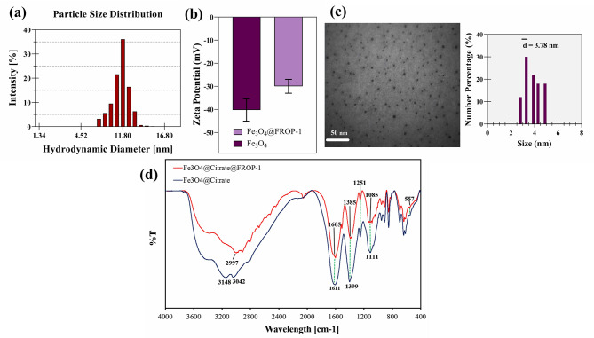
Results: In this study, the synthesis of water-soluble ultrasmall Fe3O4 NPs was performed by the co-precipitation method. XRD, TEM, and VSM analyses showed the formation of the Fe3O4 NPs with an average size of about 3.78 ± 0.2 nm. FT-IR spectroscopy approved the conjugation of the FROP-1 peptide with the Fe3O4 NPs. The synthesized Fe3O4@FROP-1 NPs showed good biocompatibility, and the high r1 relaxivity and r2/r1, respectively, were 2.608 mM- 1S- 1 and 1.18. The biocompatibility of the Fe3O4 and Fe3O4@FROP-1 NPs on the MCF-7, SKBR-3, MDA-MB-231, and MCF-10 cell lines was determined using cytotoxicity analysis. The specific targeting effect on the cells was verified by in vitro cellular uptake and cell MR imaging.
Conclusion: It was found that the contrast intensity of the Fe3O4@FROP-1 nanoprobe increases as Fe concentration increases. Cellular uptake of the Fe3O4 and Fe3O4@FROP-1 NPs was quantified using ICP-MS. The synthesized NPs had better imaging performance than Dotarem (gadoterate meglumine). The findings showed that Fe3O4@FROP-1 NPs have potential utility as a specific and targeted T1-weighted contrast agent in breast cancer MR imaging.

 求助内容:
求助内容: 应助结果提醒方式:


