Zackery Oakey, Yağmur Seda Yeşiltaş, Emily C Zabor, Arun D Singh
{"title":"不确定脉络膜黑色素细胞肿瘤的生长:到恶性转化的时间。","authors":"Zackery Oakey, Yağmur Seda Yeşiltaş, Emily C Zabor, Arun D Singh","doi":"10.4103/tjo.TJO-D-24-00138","DOIUrl":null,"url":null,"abstract":"<p><p>Indeterminate choroidal melanocytic tumors may exhibit growth under observation, but the clinical significance of early versus late growth remains unclear. In this review we aim to explore the temporal profile of growth to detect benign and malignant subsets. There was no specific set of tumor dimensions or characteristics applied for exclusion or inclusion except that all included tumors had to be described as indeterminate or suspicious nevus and observed for growth by the authors. Nine published studies (1977-2021) that included 8766 patients were reviewed. The primary outcomes were time to event (growth) and its correlation with the growth rate of small choroidal melanoma. The studies were largely retrospective and had differing inclusion criteria: largest basal diameter of 7.5-24 mm (mean, 14.83 mm) and maximum height of 2-6.7 mm (mean, 4.19 mm). Most defined growth by a change in height of >0.3 mm or base of >0.5. Among 8606 tumors followed, 478 (5.6%) showed growth over 5 years, with growth rates in individual studies ranging from 2% to 55% (mean: 19.0%). The highest event rate occurred in the 1st year (range: 0.7%-15%; mean: 6.5%), stabilizing to 0%-5% (mean: 1.5%) by year 5. The majority (range: 52%-83%; mean: 67%) were observed to grow in the initial 2 years (early growth). Over subsequent years (late growth), the proportion of growing tumors reduced and stabilized by years 4 and 5 to a mean of 6.1% and 8.3%, respectively. Time-to-event analysis indicates the heterogeneity of IMTs. Tumors growing in the first 2 years (early growth) may be melanoma in the evolution and their characteristics previously identified as the risk factors for growth may be interpreted as the factors predictive or diagnostic of small choroidal melanoma. In contrast, tumors demonstrating growth in subsequent years (late growth), after documented stability, may represent malignant transformation of a choroidal nevus.</p>","PeriodicalId":44978,"journal":{"name":"Taiwan Journal of Ophthalmology","volume":"15 1","pages":"73-78"},"PeriodicalIF":1.2000,"publicationDate":"2025-02-06","publicationTypes":"Journal Article","fieldsOfStudy":null,"isOpenAccess":false,"openAccessPdf":"https://www.ncbi.nlm.nih.gov/pmc/articles/PMC11981566/pdf/","citationCount":"0","resultStr":"{\"title\":\"Growth of indeterminate choroidal melanocytic tumors: Time to malignant transformation.\",\"authors\":\"Zackery Oakey, Yağmur Seda Yeşiltaş, Emily C Zabor, Arun D Singh\",\"doi\":\"10.4103/tjo.TJO-D-24-00138\",\"DOIUrl\":null,\"url\":null,\"abstract\":\"<p><p>Indeterminate choroidal melanocytic tumors may exhibit growth under observation, but the clinical significance of early versus late growth remains unclear. In this review we aim to explore the temporal profile of growth to detect benign and malignant subsets. There was no specific set of tumor dimensions or characteristics applied for exclusion or inclusion except that all included tumors had to be described as indeterminate or suspicious nevus and observed for growth by the authors. Nine published studies (1977-2021) that included 8766 patients were reviewed. The primary outcomes were time to event (growth) and its correlation with the growth rate of small choroidal melanoma. The studies were largely retrospective and had differing inclusion criteria: largest basal diameter of 7.5-24 mm (mean, 14.83 mm) and maximum height of 2-6.7 mm (mean, 4.19 mm). Most defined growth by a change in height of >0.3 mm or base of >0.5. Among 8606 tumors followed, 478 (5.6%) showed growth over 5 years, with growth rates in individual studies ranging from 2% to 55% (mean: 19.0%). The highest event rate occurred in the 1st year (range: 0.7%-15%; mean: 6.5%), stabilizing to 0%-5% (mean: 1.5%) by year 5. The majority (range: 52%-83%; mean: 67%) were observed to grow in the initial 2 years (early growth). Over subsequent years (late growth), the proportion of growing tumors reduced and stabilized by years 4 and 5 to a mean of 6.1% and 8.3%, respectively. Time-to-event analysis indicates the heterogeneity of IMTs. Tumors growing in the first 2 years (early growth) may be melanoma in the evolution and their characteristics previously identified as the risk factors for growth may be interpreted as the factors predictive or diagnostic of small choroidal melanoma. In contrast, tumors demonstrating growth in subsequent years (late growth), after documented stability, may represent malignant transformation of a choroidal nevus.</p>\",\"PeriodicalId\":44978,\"journal\":{\"name\":\"Taiwan Journal of Ophthalmology\",\"volume\":\"15 1\",\"pages\":\"73-78\"},\"PeriodicalIF\":1.2000,\"publicationDate\":\"2025-02-06\",\"publicationTypes\":\"Journal Article\",\"fieldsOfStudy\":null,\"isOpenAccess\":false,\"openAccessPdf\":\"https://www.ncbi.nlm.nih.gov/pmc/articles/PMC11981566/pdf/\",\"citationCount\":\"0\",\"resultStr\":null,\"platform\":\"Semanticscholar\",\"paperid\":null,\"PeriodicalName\":\"Taiwan Journal of Ophthalmology\",\"FirstCategoryId\":\"1085\",\"ListUrlMain\":\"https://doi.org/10.4103/tjo.TJO-D-24-00138\",\"RegionNum\":0,\"RegionCategory\":null,\"ArticlePicture\":[],\"TitleCN\":null,\"AbstractTextCN\":null,\"PMCID\":null,\"EPubDate\":\"2025/1/1 0:00:00\",\"PubModel\":\"eCollection\",\"JCR\":\"Q4\",\"JCRName\":\"OPHTHALMOLOGY\",\"Score\":null,\"Total\":0}","platform":"Semanticscholar","paperid":null,"PeriodicalName":"Taiwan Journal of Ophthalmology","FirstCategoryId":"1085","ListUrlMain":"https://doi.org/10.4103/tjo.TJO-D-24-00138","RegionNum":0,"RegionCategory":null,"ArticlePicture":[],"TitleCN":null,"AbstractTextCN":null,"PMCID":null,"EPubDate":"2025/1/1 0:00:00","PubModel":"eCollection","JCR":"Q4","JCRName":"OPHTHALMOLOGY","Score":null,"Total":0}
Growth of indeterminate choroidal melanocytic tumors: Time to malignant transformation.
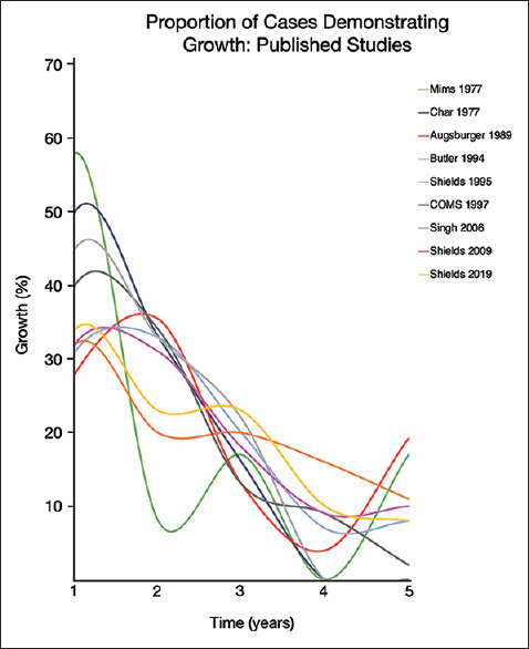
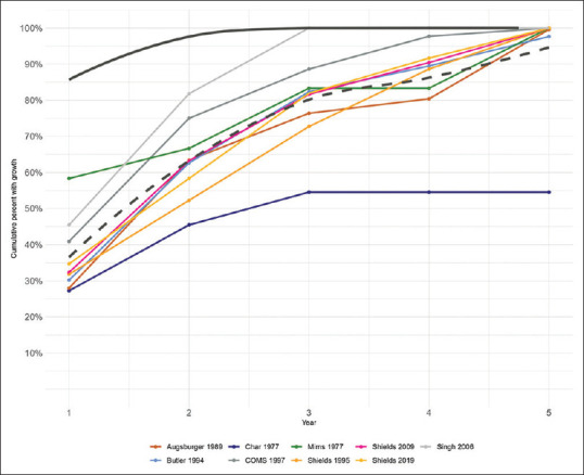
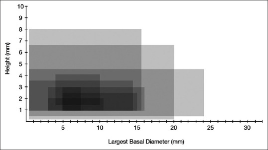
Indeterminate choroidal melanocytic tumors may exhibit growth under observation, but the clinical significance of early versus late growth remains unclear. In this review we aim to explore the temporal profile of growth to detect benign and malignant subsets. There was no specific set of tumor dimensions or characteristics applied for exclusion or inclusion except that all included tumors had to be described as indeterminate or suspicious nevus and observed for growth by the authors. Nine published studies (1977-2021) that included 8766 patients were reviewed. The primary outcomes were time to event (growth) and its correlation with the growth rate of small choroidal melanoma. The studies were largely retrospective and had differing inclusion criteria: largest basal diameter of 7.5-24 mm (mean, 14.83 mm) and maximum height of 2-6.7 mm (mean, 4.19 mm). Most defined growth by a change in height of >0.3 mm or base of >0.5. Among 8606 tumors followed, 478 (5.6%) showed growth over 5 years, with growth rates in individual studies ranging from 2% to 55% (mean: 19.0%). The highest event rate occurred in the 1st year (range: 0.7%-15%; mean: 6.5%), stabilizing to 0%-5% (mean: 1.5%) by year 5. The majority (range: 52%-83%; mean: 67%) were observed to grow in the initial 2 years (early growth). Over subsequent years (late growth), the proportion of growing tumors reduced and stabilized by years 4 and 5 to a mean of 6.1% and 8.3%, respectively. Time-to-event analysis indicates the heterogeneity of IMTs. Tumors growing in the first 2 years (early growth) may be melanoma in the evolution and their characteristics previously identified as the risk factors for growth may be interpreted as the factors predictive or diagnostic of small choroidal melanoma. In contrast, tumors demonstrating growth in subsequent years (late growth), after documented stability, may represent malignant transformation of a choroidal nevus.

 求助内容:
求助内容: 应助结果提醒方式:


