{"title":"计算机断层成像的临床实践进展及其对后纵韧带颈椎骨化手术策略的影响:系统综述。","authors":"Wongthawat Liawrungrueang, Jong-Beom Park, Watcharaporn Cholamjiak, Sompoom Sunpaweravong, Peem Sarasombath, Chaiyapruk Pundee","doi":"10.1177/20503121251331795","DOIUrl":null,"url":null,"abstract":"<p><strong>Objectives: </strong>This systematic review examines advancements in computed tomography imaging-based classification systems and their implications for surgical decision-making in managing cervical ossification of the posterior longitudinal ligament.</p><p><strong>Methods: </strong>This study is a systematic review. A comprehensive search of PubMed, MEDLINE, and Scopus databases identified relevant studies published from January 2010 to July 2024. The search utilized keywords including \"ossification of the posterior longitudinal ligament,\" \"cervical,\" \"spine,\" \"computed tomography,\" and \"classification.\" Studies meeting predefined inclusion criteria focused on computed tomography imaging for diagnosing and surgically managing cervical ossification of the posterior longitudinal ligament. The study adhered to Preferred Reporting Items for Systematic Reviews and Meta-Analyses guidelines, and the ROBINS-I tool was used for risk of bias assessment.</p><p><strong>Results: </strong>Sixteen studies were included, demonstrating that computed tomography imaging enhances diagnostic precision and classification reliability for cervical ossification of the posterior longitudinal ligament. Comparative analysis across studies revealed consistent trends in computed tomography-based classification improving surgical decision-making, particularly influencing anterior approaches such as anterior controllable antedisplacement and fusion. However, moderate to severe risks of bias were identified in some studies, primarily due to confounding variables and deviations from intended interventions. Additionally, computed tomography imaging's role in prevalence studies has been expanded by incorporating, which highlights its epidemiological significance. The review also discusses the disadvantages of computed tomography, including radiation exposure and cost implications.</p><p><strong>Conclusions: </strong>Computed tomography imaging is a crucial modality for diagnosing and managing cervical ossification of the posterior longitudinal ligament, offering superior lesion classification and guiding surgical decision-making. Future research should refine classification systems and integrate multimodal imaging approaches to enhance diagnostic and therapeutic precision.</p>","PeriodicalId":21398,"journal":{"name":"SAGE Open Medicine","volume":"13 ","pages":"20503121251331795"},"PeriodicalIF":2.1000,"publicationDate":"2025-04-12","publicationTypes":"Journal Article","fieldsOfStudy":null,"isOpenAccess":false,"openAccessPdf":"https://www.ncbi.nlm.nih.gov/pmc/articles/PMC12033476/pdf/","citationCount":"0","resultStr":"{\"title\":\"Advancements in clinical practices of computed tomography imaging and its implications on surgical strategies for the management of cervical ossification of the posterior longitudinal ligament: A systematic review.\",\"authors\":\"Wongthawat Liawrungrueang, Jong-Beom Park, Watcharaporn Cholamjiak, Sompoom Sunpaweravong, Peem Sarasombath, Chaiyapruk Pundee\",\"doi\":\"10.1177/20503121251331795\",\"DOIUrl\":null,\"url\":null,\"abstract\":\"<p><strong>Objectives: </strong>This systematic review examines advancements in computed tomography imaging-based classification systems and their implications for surgical decision-making in managing cervical ossification of the posterior longitudinal ligament.</p><p><strong>Methods: </strong>This study is a systematic review. A comprehensive search of PubMed, MEDLINE, and Scopus databases identified relevant studies published from January 2010 to July 2024. The search utilized keywords including \\\"ossification of the posterior longitudinal ligament,\\\" \\\"cervical,\\\" \\\"spine,\\\" \\\"computed tomography,\\\" and \\\"classification.\\\" Studies meeting predefined inclusion criteria focused on computed tomography imaging for diagnosing and surgically managing cervical ossification of the posterior longitudinal ligament. The study adhered to Preferred Reporting Items for Systematic Reviews and Meta-Analyses guidelines, and the ROBINS-I tool was used for risk of bias assessment.</p><p><strong>Results: </strong>Sixteen studies were included, demonstrating that computed tomography imaging enhances diagnostic precision and classification reliability for cervical ossification of the posterior longitudinal ligament. Comparative analysis across studies revealed consistent trends in computed tomography-based classification improving surgical decision-making, particularly influencing anterior approaches such as anterior controllable antedisplacement and fusion. However, moderate to severe risks of bias were identified in some studies, primarily due to confounding variables and deviations from intended interventions. Additionally, computed tomography imaging's role in prevalence studies has been expanded by incorporating, which highlights its epidemiological significance. The review also discusses the disadvantages of computed tomography, including radiation exposure and cost implications.</p><p><strong>Conclusions: </strong>Computed tomography imaging is a crucial modality for diagnosing and managing cervical ossification of the posterior longitudinal ligament, offering superior lesion classification and guiding surgical decision-making. Future research should refine classification systems and integrate multimodal imaging approaches to enhance diagnostic and therapeutic precision.</p>\",\"PeriodicalId\":21398,\"journal\":{\"name\":\"SAGE Open Medicine\",\"volume\":\"13 \",\"pages\":\"20503121251331795\"},\"PeriodicalIF\":2.1000,\"publicationDate\":\"2025-04-12\",\"publicationTypes\":\"Journal Article\",\"fieldsOfStudy\":null,\"isOpenAccess\":false,\"openAccessPdf\":\"https://www.ncbi.nlm.nih.gov/pmc/articles/PMC12033476/pdf/\",\"citationCount\":\"0\",\"resultStr\":null,\"platform\":\"Semanticscholar\",\"paperid\":null,\"PeriodicalName\":\"SAGE Open Medicine\",\"FirstCategoryId\":\"1085\",\"ListUrlMain\":\"https://doi.org/10.1177/20503121251331795\",\"RegionNum\":0,\"RegionCategory\":null,\"ArticlePicture\":[],\"TitleCN\":null,\"AbstractTextCN\":null,\"PMCID\":null,\"EPubDate\":\"2025/1/1 0:00:00\",\"PubModel\":\"eCollection\",\"JCR\":\"Q2\",\"JCRName\":\"MEDICINE, GENERAL & INTERNAL\",\"Score\":null,\"Total\":0}","platform":"Semanticscholar","paperid":null,"PeriodicalName":"SAGE Open Medicine","FirstCategoryId":"1085","ListUrlMain":"https://doi.org/10.1177/20503121251331795","RegionNum":0,"RegionCategory":null,"ArticlePicture":[],"TitleCN":null,"AbstractTextCN":null,"PMCID":null,"EPubDate":"2025/1/1 0:00:00","PubModel":"eCollection","JCR":"Q2","JCRName":"MEDICINE, GENERAL & INTERNAL","Score":null,"Total":0}
Advancements in clinical practices of computed tomography imaging and its implications on surgical strategies for the management of cervical ossification of the posterior longitudinal ligament: A systematic review.
Objectives: This systematic review examines advancements in computed tomography imaging-based classification systems and their implications for surgical decision-making in managing cervical ossification of the posterior longitudinal ligament.
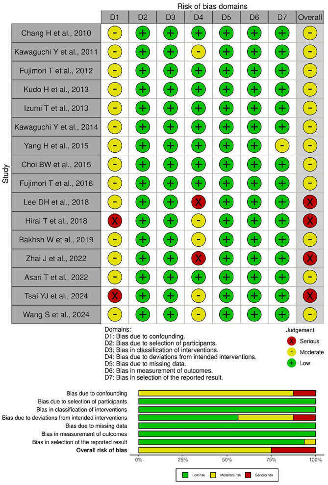
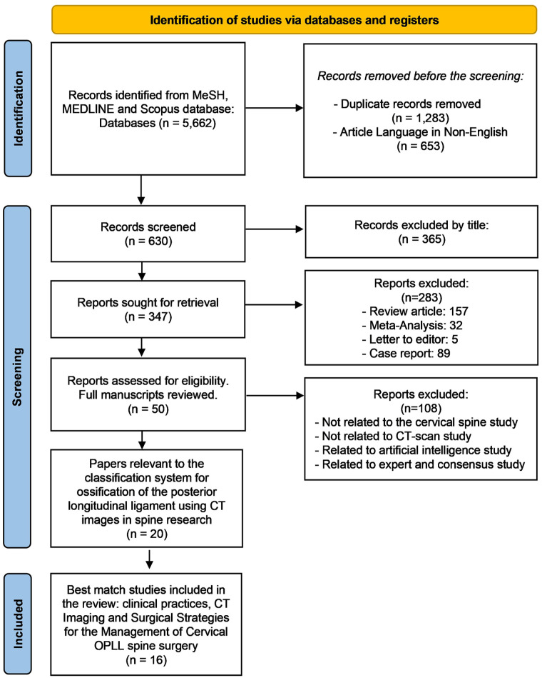
Methods: This study is a systematic review. A comprehensive search of PubMed, MEDLINE, and Scopus databases identified relevant studies published from January 2010 to July 2024. The search utilized keywords including "ossification of the posterior longitudinal ligament," "cervical," "spine," "computed tomography," and "classification." Studies meeting predefined inclusion criteria focused on computed tomography imaging for diagnosing and surgically managing cervical ossification of the posterior longitudinal ligament. The study adhered to Preferred Reporting Items for Systematic Reviews and Meta-Analyses guidelines, and the ROBINS-I tool was used for risk of bias assessment.
Results: Sixteen studies were included, demonstrating that computed tomography imaging enhances diagnostic precision and classification reliability for cervical ossification of the posterior longitudinal ligament. Comparative analysis across studies revealed consistent trends in computed tomography-based classification improving surgical decision-making, particularly influencing anterior approaches such as anterior controllable antedisplacement and fusion. However, moderate to severe risks of bias were identified in some studies, primarily due to confounding variables and deviations from intended interventions. Additionally, computed tomography imaging's role in prevalence studies has been expanded by incorporating, which highlights its epidemiological significance. The review also discusses the disadvantages of computed tomography, including radiation exposure and cost implications.
Conclusions: Computed tomography imaging is a crucial modality for diagnosing and managing cervical ossification of the posterior longitudinal ligament, offering superior lesion classification and guiding surgical decision-making. Future research should refine classification systems and integrate multimodal imaging approaches to enhance diagnostic and therapeutic precision.


 求助内容:
求助内容: 应助结果提醒方式:


