{"title":"新型幼年犬骨化性咽皮样囊肿的计算机断层特征分析。","authors":"Kathleen Kalphat-Losego, Robson F Giglio","doi":"10.1111/vru.70036","DOIUrl":null,"url":null,"abstract":"<p><p>A 6-month-old, intact female Boxer presented with ongoing dyspnea, nasal congestion, cardiac arrhythmias, stertor, and syncope. A CT assessment of the head, neck, thorax, and abdomen revealed a fluid to soft tissue attenuating mass with an incomplete mineralized rim centered on the soft tissue ventral to the right tympanic bulla. Histopathologically, epithelialization was consistent with a dermoid cyst. The severe degree of mass effect caused by this abnormally and undocumented ossified structure in vital cranial cervical organs causes more systemic complications than traditional dermoid cysts. CT delineated the cyst's structure and compression/occlusion of adjacent structures.</p>","PeriodicalId":23581,"journal":{"name":"Veterinary Radiology & Ultrasound","volume":"66 3","pages":"e70036"},"PeriodicalIF":1.5000,"publicationDate":"2025-05-01","publicationTypes":"Journal Article","fieldsOfStudy":null,"isOpenAccess":false,"openAccessPdf":"https://www.ncbi.nlm.nih.gov/pmc/articles/PMC12041625/pdf/","citationCount":"0","resultStr":"{\"title\":\"Computed Tomographic Characteristics Associated With Novel Case of Ossified Pharyngeal Dermoid Cyst in Juvenile Canine.\",\"authors\":\"Kathleen Kalphat-Losego, Robson F Giglio\",\"doi\":\"10.1111/vru.70036\",\"DOIUrl\":null,\"url\":null,\"abstract\":\"<p><p>A 6-month-old, intact female Boxer presented with ongoing dyspnea, nasal congestion, cardiac arrhythmias, stertor, and syncope. A CT assessment of the head, neck, thorax, and abdomen revealed a fluid to soft tissue attenuating mass with an incomplete mineralized rim centered on the soft tissue ventral to the right tympanic bulla. Histopathologically, epithelialization was consistent with a dermoid cyst. The severe degree of mass effect caused by this abnormally and undocumented ossified structure in vital cranial cervical organs causes more systemic complications than traditional dermoid cysts. CT delineated the cyst's structure and compression/occlusion of adjacent structures.</p>\",\"PeriodicalId\":23581,\"journal\":{\"name\":\"Veterinary Radiology & Ultrasound\",\"volume\":\"66 3\",\"pages\":\"e70036\"},\"PeriodicalIF\":1.5000,\"publicationDate\":\"2025-05-01\",\"publicationTypes\":\"Journal Article\",\"fieldsOfStudy\":null,\"isOpenAccess\":false,\"openAccessPdf\":\"https://www.ncbi.nlm.nih.gov/pmc/articles/PMC12041625/pdf/\",\"citationCount\":\"0\",\"resultStr\":null,\"platform\":\"Semanticscholar\",\"paperid\":null,\"PeriodicalName\":\"Veterinary Radiology & Ultrasound\",\"FirstCategoryId\":\"97\",\"ListUrlMain\":\"https://doi.org/10.1111/vru.70036\",\"RegionNum\":2,\"RegionCategory\":\"农林科学\",\"ArticlePicture\":[],\"TitleCN\":null,\"AbstractTextCN\":null,\"PMCID\":null,\"EPubDate\":\"\",\"PubModel\":\"\",\"JCR\":\"Q2\",\"JCRName\":\"VETERINARY SCIENCES\",\"Score\":null,\"Total\":0}","platform":"Semanticscholar","paperid":null,"PeriodicalName":"Veterinary Radiology & Ultrasound","FirstCategoryId":"97","ListUrlMain":"https://doi.org/10.1111/vru.70036","RegionNum":2,"RegionCategory":"农林科学","ArticlePicture":[],"TitleCN":null,"AbstractTextCN":null,"PMCID":null,"EPubDate":"","PubModel":"","JCR":"Q2","JCRName":"VETERINARY SCIENCES","Score":null,"Total":0}
Computed Tomographic Characteristics Associated With Novel Case of Ossified Pharyngeal Dermoid Cyst in Juvenile Canine.
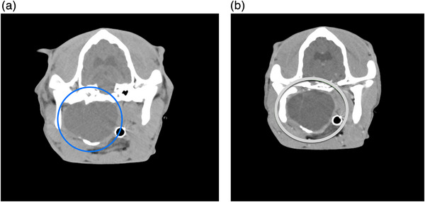
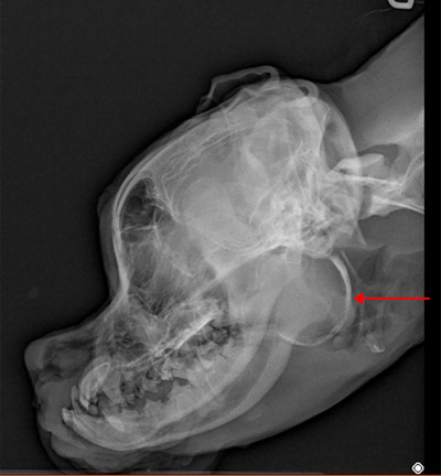
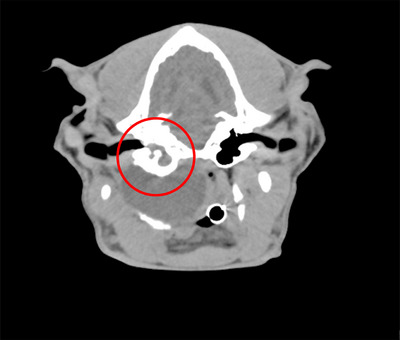
A 6-month-old, intact female Boxer presented with ongoing dyspnea, nasal congestion, cardiac arrhythmias, stertor, and syncope. A CT assessment of the head, neck, thorax, and abdomen revealed a fluid to soft tissue attenuating mass with an incomplete mineralized rim centered on the soft tissue ventral to the right tympanic bulla. Histopathologically, epithelialization was consistent with a dermoid cyst. The severe degree of mass effect caused by this abnormally and undocumented ossified structure in vital cranial cervical organs causes more systemic complications than traditional dermoid cysts. CT delineated the cyst's structure and compression/occlusion of adjacent structures.
期刊介绍:
Veterinary Radiology & Ultrasound is a bimonthly, international, peer-reviewed, research journal devoted to the fields of veterinary diagnostic imaging and radiation oncology. Established in 1958, it is owned by the American College of Veterinary Radiology and is also the official journal for six affiliate veterinary organizations. Veterinary Radiology & Ultrasound is represented on the International Committee of Medical Journal Editors, World Association of Medical Editors, and Committee on Publication Ethics.
The mission of Veterinary Radiology & Ultrasound is to serve as a leading resource for high quality articles that advance scientific knowledge and standards of clinical practice in the areas of veterinary diagnostic radiology, computed tomography, magnetic resonance imaging, ultrasonography, nuclear imaging, radiation oncology, and interventional radiology. Manuscript types include original investigations, imaging diagnosis reports, review articles, editorials and letters to the Editor. Acceptance criteria include originality, significance, quality, reader interest, composition and adherence to author guidelines.

 求助内容:
求助内容: 应助结果提醒方式:


