Min-Bo Zhu, Bei-Lei Chen, Meng Cen, Li-Ping Chen, Zheng Shi
{"title":"子宫内膜腺显像与细泡检测评价子宫内膜容受性的比较分析。","authors":"Min-Bo Zhu, Bei-Lei Chen, Meng Cen, Li-Ping Chen, Zheng Shi","doi":"10.1186/s12958-025-01395-y","DOIUrl":null,"url":null,"abstract":"<p><strong>Background: </strong>This study aimed to obtain high-resolution endometrial gland images under high-definition hysteroscopy from in vitro fertilization and embryo transfer (IVF-ET) patients and perform image recognition to analyze their density and opening size. Concurrently, the number and morphology of pinopodes in the endometrial samples were observed using scanning electron microscopy (SEM). The objective was to compare the correlation between the two methods in evaluating endometrial receptivity and predicting pregnancy outcomes and to assess the consistency between the quantitative curves and pregnancy outcomes of the two methods in the cohort study.</p><p><strong>Methods: </strong>67 patients undergoing hysteroscopic surgery who had undergone controlled ovarian hyperstimulation treatment within 1-3 menstrual cycles before the surgery were selected. Hysteroscopic exploration was performed on the 3-5 days after ovulation (during the implantation window period). Endometrial images and tissues were collected under hysteroscopy. The endometrial glands were counted, the sizes of gland openings were calculated using an image recognition algorithm, and the number and morphology of endometrial pinopodes were observed through SEM. All patients underwent embryo transfer surgery within 1-3 menstrual cycles after hysteroscopy. Patients were divided into pregnancy and non-pregnancy groups based on pregnancy outcomes. The density and size of endometrial glands, as well as the number and morphology of pinopodes were compared between the two groups.</p><p><strong>Results: </strong>The endometrial gland density and the size of the endometrial gland opening in the pregnancy group were higher than that in the non-pregnancy group (P < 0.05). In contrast, both the average pinopode count per image and the developmental maturity grading of pinopodes were significantly higher in the pregnancy group compared to the non-pregnancy group.</p><p><strong>Conclusions: </strong>Both hysteroscopic endometrial gland image recognition technology and pinopode detection can effectively reflect endometrial receptivity and predict pregnancy outcomes. However, image recognition technology has clear economic and promotional advantages.</p><p><strong>Clinical trial number: </strong>Not applicable.</p>","PeriodicalId":21011,"journal":{"name":"Reproductive Biology and Endocrinology","volume":"23 1","pages":"62"},"PeriodicalIF":4.7000,"publicationDate":"2025-04-28","publicationTypes":"Journal Article","fieldsOfStudy":null,"isOpenAccess":false,"openAccessPdf":"https://www.ncbi.nlm.nih.gov/pmc/articles/PMC12036253/pdf/","citationCount":"0","resultStr":"{\"title\":\"Comparative analysis of endometrial gland imaging and pinopode detection for assessing endometrial receptivity.\",\"authors\":\"Min-Bo Zhu, Bei-Lei Chen, Meng Cen, Li-Ping Chen, Zheng Shi\",\"doi\":\"10.1186/s12958-025-01395-y\",\"DOIUrl\":null,\"url\":null,\"abstract\":\"<p><strong>Background: </strong>This study aimed to obtain high-resolution endometrial gland images under high-definition hysteroscopy from in vitro fertilization and embryo transfer (IVF-ET) patients and perform image recognition to analyze their density and opening size. Concurrently, the number and morphology of pinopodes in the endometrial samples were observed using scanning electron microscopy (SEM). The objective was to compare the correlation between the two methods in evaluating endometrial receptivity and predicting pregnancy outcomes and to assess the consistency between the quantitative curves and pregnancy outcomes of the two methods in the cohort study.</p><p><strong>Methods: </strong>67 patients undergoing hysteroscopic surgery who had undergone controlled ovarian hyperstimulation treatment within 1-3 menstrual cycles before the surgery were selected. Hysteroscopic exploration was performed on the 3-5 days after ovulation (during the implantation window period). Endometrial images and tissues were collected under hysteroscopy. The endometrial glands were counted, the sizes of gland openings were calculated using an image recognition algorithm, and the number and morphology of endometrial pinopodes were observed through SEM. All patients underwent embryo transfer surgery within 1-3 menstrual cycles after hysteroscopy. Patients were divided into pregnancy and non-pregnancy groups based on pregnancy outcomes. The density and size of endometrial glands, as well as the number and morphology of pinopodes were compared between the two groups.</p><p><strong>Results: </strong>The endometrial gland density and the size of the endometrial gland opening in the pregnancy group were higher than that in the non-pregnancy group (P < 0.05). In contrast, both the average pinopode count per image and the developmental maturity grading of pinopodes were significantly higher in the pregnancy group compared to the non-pregnancy group.</p><p><strong>Conclusions: </strong>Both hysteroscopic endometrial gland image recognition technology and pinopode detection can effectively reflect endometrial receptivity and predict pregnancy outcomes. However, image recognition technology has clear economic and promotional advantages.</p><p><strong>Clinical trial number: </strong>Not applicable.</p>\",\"PeriodicalId\":21011,\"journal\":{\"name\":\"Reproductive Biology and Endocrinology\",\"volume\":\"23 1\",\"pages\":\"62\"},\"PeriodicalIF\":4.7000,\"publicationDate\":\"2025-04-28\",\"publicationTypes\":\"Journal Article\",\"fieldsOfStudy\":null,\"isOpenAccess\":false,\"openAccessPdf\":\"https://www.ncbi.nlm.nih.gov/pmc/articles/PMC12036253/pdf/\",\"citationCount\":\"0\",\"resultStr\":null,\"platform\":\"Semanticscholar\",\"paperid\":null,\"PeriodicalName\":\"Reproductive Biology and Endocrinology\",\"FirstCategoryId\":\"3\",\"ListUrlMain\":\"https://doi.org/10.1186/s12958-025-01395-y\",\"RegionNum\":2,\"RegionCategory\":\"医学\",\"ArticlePicture\":[],\"TitleCN\":null,\"AbstractTextCN\":null,\"PMCID\":null,\"EPubDate\":\"\",\"PubModel\":\"\",\"JCR\":\"Q1\",\"JCRName\":\"ENDOCRINOLOGY & METABOLISM\",\"Score\":null,\"Total\":0}","platform":"Semanticscholar","paperid":null,"PeriodicalName":"Reproductive Biology and Endocrinology","FirstCategoryId":"3","ListUrlMain":"https://doi.org/10.1186/s12958-025-01395-y","RegionNum":2,"RegionCategory":"医学","ArticlePicture":[],"TitleCN":null,"AbstractTextCN":null,"PMCID":null,"EPubDate":"","PubModel":"","JCR":"Q1","JCRName":"ENDOCRINOLOGY & METABOLISM","Score":null,"Total":0}
Comparative analysis of endometrial gland imaging and pinopode detection for assessing endometrial receptivity.
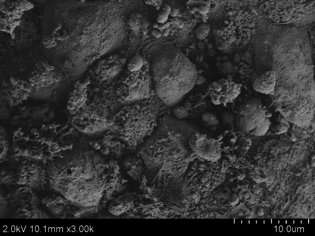
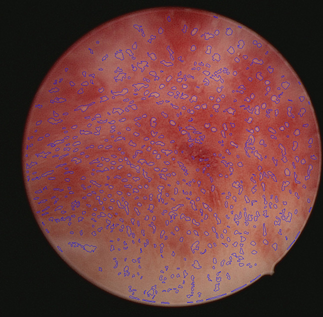
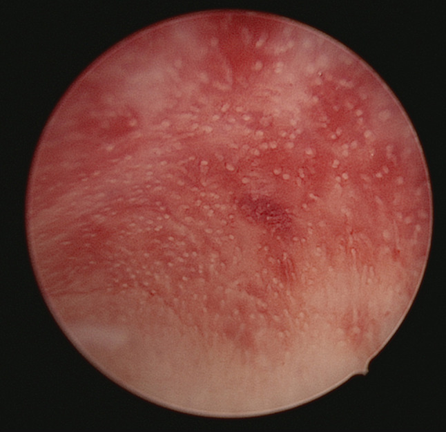
Background: This study aimed to obtain high-resolution endometrial gland images under high-definition hysteroscopy from in vitro fertilization and embryo transfer (IVF-ET) patients and perform image recognition to analyze their density and opening size. Concurrently, the number and morphology of pinopodes in the endometrial samples were observed using scanning electron microscopy (SEM). The objective was to compare the correlation between the two methods in evaluating endometrial receptivity and predicting pregnancy outcomes and to assess the consistency between the quantitative curves and pregnancy outcomes of the two methods in the cohort study.
Methods: 67 patients undergoing hysteroscopic surgery who had undergone controlled ovarian hyperstimulation treatment within 1-3 menstrual cycles before the surgery were selected. Hysteroscopic exploration was performed on the 3-5 days after ovulation (during the implantation window period). Endometrial images and tissues were collected under hysteroscopy. The endometrial glands were counted, the sizes of gland openings were calculated using an image recognition algorithm, and the number and morphology of endometrial pinopodes were observed through SEM. All patients underwent embryo transfer surgery within 1-3 menstrual cycles after hysteroscopy. Patients were divided into pregnancy and non-pregnancy groups based on pregnancy outcomes. The density and size of endometrial glands, as well as the number and morphology of pinopodes were compared between the two groups.
Results: The endometrial gland density and the size of the endometrial gland opening in the pregnancy group were higher than that in the non-pregnancy group (P < 0.05). In contrast, both the average pinopode count per image and the developmental maturity grading of pinopodes were significantly higher in the pregnancy group compared to the non-pregnancy group.
Conclusions: Both hysteroscopic endometrial gland image recognition technology and pinopode detection can effectively reflect endometrial receptivity and predict pregnancy outcomes. However, image recognition technology has clear economic and promotional advantages.
期刊介绍:
Reproductive Biology and Endocrinology publishes and disseminates high-quality results from excellent research in the reproductive sciences.
The journal publishes on topics covering gametogenesis, fertilization, early embryonic development, embryo-uterus interaction, reproductive development, pregnancy, uterine biology, endocrinology of reproduction, control of reproduction, reproductive immunology, neuroendocrinology, and veterinary and human reproductive medicine, including all vertebrate species.
 求助内容:
求助内容: 应助结果提醒方式:


