Hyoung Soo Choi, Chae-Yong Kim, Byung Se Choi, Seung Hyuck Jeon, In Ah Kim, Joo-Young Kim, Kyu Sang Lee, Gheeyoung Choe
{"title":"辐射致小脑海绵状畸形2例临床分析。","authors":"Hyoung Soo Choi, Chae-Yong Kim, Byung Se Choi, Seung Hyuck Jeon, In Ah Kim, Joo-Young Kim, Kyu Sang Lee, Gheeyoung Choe","doi":"10.14791/btrt.2025.0003","DOIUrl":null,"url":null,"abstract":"<p><p>Radiation-induced cavernous malformations (RICMs) are rare but significant late complications of high-dose radiation therapy, particularly in young survivors of brain tumors. This report presents two cases of RICMs following aggressive multimodal treatment, including surgery, chemotherapy, and radiation therapy. Case 1 was a 22-year-old male patient with medulloblastoma treated with craniospinal irradiation, tumor bed boost, and tandem autologous peripheral blood stem cell transplantation. Approximately 8 years after treatment completion, routine follow-up imaging revealed a small focal hemorrhage in the right cerebellum, consistent with an RICM. The lesion was asymptomatic and managed conservatively with regular imaging, showing spontaneous resolution over time, with a significant size reduction noted 9 years post-treatment. Case 2 describes a 32-year-old male with an intracranial germinoma treated with whole-ventricular irradiation. Three years after treatment, the patient developed a symptomatic hemorrhagic RICM near a pre-existing developmental venous anomaly. Surgical resection and Gamma Knife Surgery stabilized the lesion; however, residual symptoms, including tremors and gait disturbances, persisted, affecting the patient's daily activities. These cases illustrate the diverse clinical courses of RICMs, ranging from spontaneous resolution to the necessity of surgical intervention, and emphasize the importance of long-term surveillance and tailored management strategies for late-onset complications.</p>","PeriodicalId":72453,"journal":{"name":"Brain tumor research and treatment","volume":"13 2","pages":"58-64"},"PeriodicalIF":0.0000,"publicationDate":"2025-04-01","publicationTypes":"Journal Article","fieldsOfStudy":null,"isOpenAccess":false,"openAccessPdf":"https://www.ncbi.nlm.nih.gov/pmc/articles/PMC12070075/pdf/","citationCount":"0","resultStr":"{\"title\":\"Radiation-Induced Cavernous Malformation in the Cerebellum: Clinical Features of Two Cases.\",\"authors\":\"Hyoung Soo Choi, Chae-Yong Kim, Byung Se Choi, Seung Hyuck Jeon, In Ah Kim, Joo-Young Kim, Kyu Sang Lee, Gheeyoung Choe\",\"doi\":\"10.14791/btrt.2025.0003\",\"DOIUrl\":null,\"url\":null,\"abstract\":\"<p><p>Radiation-induced cavernous malformations (RICMs) are rare but significant late complications of high-dose radiation therapy, particularly in young survivors of brain tumors. This report presents two cases of RICMs following aggressive multimodal treatment, including surgery, chemotherapy, and radiation therapy. Case 1 was a 22-year-old male patient with medulloblastoma treated with craniospinal irradiation, tumor bed boost, and tandem autologous peripheral blood stem cell transplantation. Approximately 8 years after treatment completion, routine follow-up imaging revealed a small focal hemorrhage in the right cerebellum, consistent with an RICM. The lesion was asymptomatic and managed conservatively with regular imaging, showing spontaneous resolution over time, with a significant size reduction noted 9 years post-treatment. Case 2 describes a 32-year-old male with an intracranial germinoma treated with whole-ventricular irradiation. Three years after treatment, the patient developed a symptomatic hemorrhagic RICM near a pre-existing developmental venous anomaly. Surgical resection and Gamma Knife Surgery stabilized the lesion; however, residual symptoms, including tremors and gait disturbances, persisted, affecting the patient's daily activities. These cases illustrate the diverse clinical courses of RICMs, ranging from spontaneous resolution to the necessity of surgical intervention, and emphasize the importance of long-term surveillance and tailored management strategies for late-onset complications.</p>\",\"PeriodicalId\":72453,\"journal\":{\"name\":\"Brain tumor research and treatment\",\"volume\":\"13 2\",\"pages\":\"58-64\"},\"PeriodicalIF\":0.0000,\"publicationDate\":\"2025-04-01\",\"publicationTypes\":\"Journal Article\",\"fieldsOfStudy\":null,\"isOpenAccess\":false,\"openAccessPdf\":\"https://www.ncbi.nlm.nih.gov/pmc/articles/PMC12070075/pdf/\",\"citationCount\":\"0\",\"resultStr\":null,\"platform\":\"Semanticscholar\",\"paperid\":null,\"PeriodicalName\":\"Brain tumor research and treatment\",\"FirstCategoryId\":\"1085\",\"ListUrlMain\":\"https://doi.org/10.14791/btrt.2025.0003\",\"RegionNum\":0,\"RegionCategory\":null,\"ArticlePicture\":[],\"TitleCN\":null,\"AbstractTextCN\":null,\"PMCID\":null,\"EPubDate\":\"\",\"PubModel\":\"\",\"JCR\":\"\",\"JCRName\":\"\",\"Score\":null,\"Total\":0}","platform":"Semanticscholar","paperid":null,"PeriodicalName":"Brain tumor research and treatment","FirstCategoryId":"1085","ListUrlMain":"https://doi.org/10.14791/btrt.2025.0003","RegionNum":0,"RegionCategory":null,"ArticlePicture":[],"TitleCN":null,"AbstractTextCN":null,"PMCID":null,"EPubDate":"","PubModel":"","JCR":"","JCRName":"","Score":null,"Total":0}
Radiation-Induced Cavernous Malformation in the Cerebellum: Clinical Features of Two Cases.
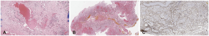
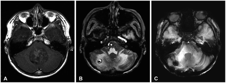
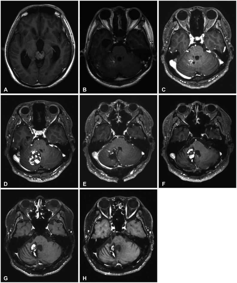
Radiation-induced cavernous malformations (RICMs) are rare but significant late complications of high-dose radiation therapy, particularly in young survivors of brain tumors. This report presents two cases of RICMs following aggressive multimodal treatment, including surgery, chemotherapy, and radiation therapy. Case 1 was a 22-year-old male patient with medulloblastoma treated with craniospinal irradiation, tumor bed boost, and tandem autologous peripheral blood stem cell transplantation. Approximately 8 years after treatment completion, routine follow-up imaging revealed a small focal hemorrhage in the right cerebellum, consistent with an RICM. The lesion was asymptomatic and managed conservatively with regular imaging, showing spontaneous resolution over time, with a significant size reduction noted 9 years post-treatment. Case 2 describes a 32-year-old male with an intracranial germinoma treated with whole-ventricular irradiation. Three years after treatment, the patient developed a symptomatic hemorrhagic RICM near a pre-existing developmental venous anomaly. Surgical resection and Gamma Knife Surgery stabilized the lesion; however, residual symptoms, including tremors and gait disturbances, persisted, affecting the patient's daily activities. These cases illustrate the diverse clinical courses of RICMs, ranging from spontaneous resolution to the necessity of surgical intervention, and emphasize the importance of long-term surveillance and tailored management strategies for late-onset complications.

 求助内容:
求助内容: 应助结果提醒方式:


