Hannes Engel, Andrea Nedelcu, Robert Grimm, Heinrich von Busch, August Sigle, Tobias Krauss, Christopher L Schlett, Jakob Weiss, Matthias Benndorf, Benedict Oerther
{"title":"一种全自动AI算法在疑似前列腺癌患者病变检测及PI-RADS分类中的诊断性能","authors":"Hannes Engel, Andrea Nedelcu, Robert Grimm, Heinrich von Busch, August Sigle, Tobias Krauss, Christopher L Schlett, Jakob Weiss, Matthias Benndorf, Benedict Oerther","doi":"10.1007/s11547-025-02003-0","DOIUrl":null,"url":null,"abstract":"<p><strong>Purpose: </strong>To evaluate the diagnostic performance of a fully automated, commercially available AI algorithm for detecting prostate cancer and classifying lesions according to PI-RADS.</p><p><strong>Material and methods: </strong>In this retrospective single-center cohort study, we included consecutive patients with suspected prostate cancer who underwent 3T MRI between May 2017 and May 2020. Histopathological ground truth was targeted transperineal ultrasound-fusion guided biopsy and extensive systematic biopsy. We compared the results of the AI algorithm to those of human readers on both the lesion and patient level and determined the diagnostic performance.</p><p><strong>Results: </strong>A total of 272 patients with 436 target lesions were evaluated. Of these patients, 135 (49.6%) had clinically significant prostate cancer (sPCa), 35 (12.9%) had clinically insignificant prostate cancer (ISUP = 1), and 102 (37.5%) were benign. On patient level, the cancer detection rates of sPCa for AI versus human readers were 11% versus 18% for PI-RADS ≤ 2, 27% versus 11% for PI-RADS 3, 54% versus 41% for PI-RADS 4, and 74% versus 92% for PI-RADS 5. The AI showed significantly higher accuracy: 74% versus 63% for PI-RADS ≥ 4 (p < 0.01) and 70% versus 52% for PI-RADS ≥ 3 (p < 0.01). Additionally, the AI correctly classified 62 patients with human reading PI-RADS ≥ 3 as true negatives.</p><p><strong>Conclusion: </strong>The AI algorithm proved to be a reliable and robust tool for lesion detection and classification. Its cancer detection rates and PI-RADS category distribution align with the results of recent meta-analyses, indicating precise risk stratification.</p>","PeriodicalId":20817,"journal":{"name":"Radiologia Medica","volume":" ","pages":"1039-1049"},"PeriodicalIF":4.8000,"publicationDate":"2025-07-01","publicationTypes":"Journal Article","fieldsOfStudy":null,"isOpenAccess":false,"openAccessPdf":"https://www.ncbi.nlm.nih.gov/pmc/articles/PMC12263473/pdf/","citationCount":"0","resultStr":"{\"title\":\"Diagnostic performance of a fully automated AI algorithm for lesion detection and PI-RADS classification in patients with suspected prostate cancer.\",\"authors\":\"Hannes Engel, Andrea Nedelcu, Robert Grimm, Heinrich von Busch, August Sigle, Tobias Krauss, Christopher L Schlett, Jakob Weiss, Matthias Benndorf, Benedict Oerther\",\"doi\":\"10.1007/s11547-025-02003-0\",\"DOIUrl\":null,\"url\":null,\"abstract\":\"<p><strong>Purpose: </strong>To evaluate the diagnostic performance of a fully automated, commercially available AI algorithm for detecting prostate cancer and classifying lesions according to PI-RADS.</p><p><strong>Material and methods: </strong>In this retrospective single-center cohort study, we included consecutive patients with suspected prostate cancer who underwent 3T MRI between May 2017 and May 2020. Histopathological ground truth was targeted transperineal ultrasound-fusion guided biopsy and extensive systematic biopsy. We compared the results of the AI algorithm to those of human readers on both the lesion and patient level and determined the diagnostic performance.</p><p><strong>Results: </strong>A total of 272 patients with 436 target lesions were evaluated. Of these patients, 135 (49.6%) had clinically significant prostate cancer (sPCa), 35 (12.9%) had clinically insignificant prostate cancer (ISUP = 1), and 102 (37.5%) were benign. On patient level, the cancer detection rates of sPCa for AI versus human readers were 11% versus 18% for PI-RADS ≤ 2, 27% versus 11% for PI-RADS 3, 54% versus 41% for PI-RADS 4, and 74% versus 92% for PI-RADS 5. The AI showed significantly higher accuracy: 74% versus 63% for PI-RADS ≥ 4 (p < 0.01) and 70% versus 52% for PI-RADS ≥ 3 (p < 0.01). Additionally, the AI correctly classified 62 patients with human reading PI-RADS ≥ 3 as true negatives.</p><p><strong>Conclusion: </strong>The AI algorithm proved to be a reliable and robust tool for lesion detection and classification. Its cancer detection rates and PI-RADS category distribution align with the results of recent meta-analyses, indicating precise risk stratification.</p>\",\"PeriodicalId\":20817,\"journal\":{\"name\":\"Radiologia Medica\",\"volume\":\" \",\"pages\":\"1039-1049\"},\"PeriodicalIF\":4.8000,\"publicationDate\":\"2025-07-01\",\"publicationTypes\":\"Journal Article\",\"fieldsOfStudy\":null,\"isOpenAccess\":false,\"openAccessPdf\":\"https://www.ncbi.nlm.nih.gov/pmc/articles/PMC12263473/pdf/\",\"citationCount\":\"0\",\"resultStr\":null,\"platform\":\"Semanticscholar\",\"paperid\":null,\"PeriodicalName\":\"Radiologia Medica\",\"FirstCategoryId\":\"3\",\"ListUrlMain\":\"https://doi.org/10.1007/s11547-025-02003-0\",\"RegionNum\":1,\"RegionCategory\":\"医学\",\"ArticlePicture\":[],\"TitleCN\":null,\"AbstractTextCN\":null,\"PMCID\":null,\"EPubDate\":\"2025/4/17 0:00:00\",\"PubModel\":\"Epub\",\"JCR\":\"Q1\",\"JCRName\":\"RADIOLOGY, NUCLEAR MEDICINE & MEDICAL IMAGING\",\"Score\":null,\"Total\":0}","platform":"Semanticscholar","paperid":null,"PeriodicalName":"Radiologia Medica","FirstCategoryId":"3","ListUrlMain":"https://doi.org/10.1007/s11547-025-02003-0","RegionNum":1,"RegionCategory":"医学","ArticlePicture":[],"TitleCN":null,"AbstractTextCN":null,"PMCID":null,"EPubDate":"2025/4/17 0:00:00","PubModel":"Epub","JCR":"Q1","JCRName":"RADIOLOGY, NUCLEAR MEDICINE & MEDICAL IMAGING","Score":null,"Total":0}
Diagnostic performance of a fully automated AI algorithm for lesion detection and PI-RADS classification in patients with suspected prostate cancer.
Purpose: To evaluate the diagnostic performance of a fully automated, commercially available AI algorithm for detecting prostate cancer and classifying lesions according to PI-RADS.
Material and methods: In this retrospective single-center cohort study, we included consecutive patients with suspected prostate cancer who underwent 3T MRI between May 2017 and May 2020. Histopathological ground truth was targeted transperineal ultrasound-fusion guided biopsy and extensive systematic biopsy. We compared the results of the AI algorithm to those of human readers on both the lesion and patient level and determined the diagnostic performance.
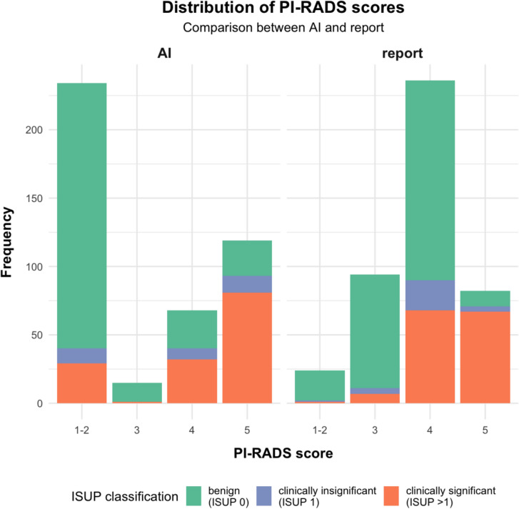
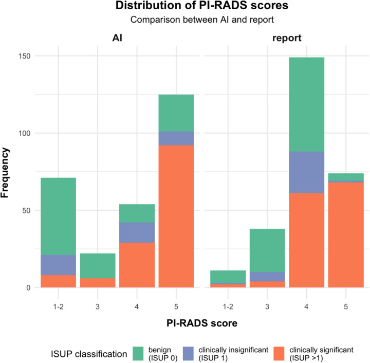
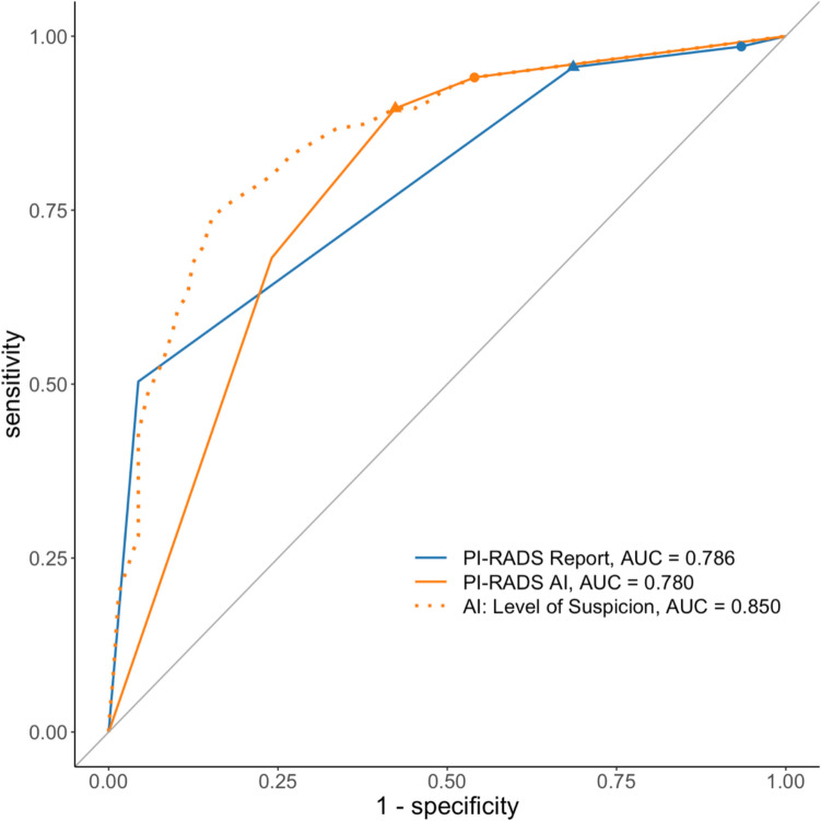
Results: A total of 272 patients with 436 target lesions were evaluated. Of these patients, 135 (49.6%) had clinically significant prostate cancer (sPCa), 35 (12.9%) had clinically insignificant prostate cancer (ISUP = 1), and 102 (37.5%) were benign. On patient level, the cancer detection rates of sPCa for AI versus human readers were 11% versus 18% for PI-RADS ≤ 2, 27% versus 11% for PI-RADS 3, 54% versus 41% for PI-RADS 4, and 74% versus 92% for PI-RADS 5. The AI showed significantly higher accuracy: 74% versus 63% for PI-RADS ≥ 4 (p < 0.01) and 70% versus 52% for PI-RADS ≥ 3 (p < 0.01). Additionally, the AI correctly classified 62 patients with human reading PI-RADS ≥ 3 as true negatives.
Conclusion: The AI algorithm proved to be a reliable and robust tool for lesion detection and classification. Its cancer detection rates and PI-RADS category distribution align with the results of recent meta-analyses, indicating precise risk stratification.
期刊介绍:
Felice Perussia founded La radiologia medica in 1914. It is a peer-reviewed journal and serves as the official journal of the Italian Society of Medical and Interventional Radiology (SIRM). The primary purpose of the journal is to disseminate information related to Radiology, especially advancements in diagnostic imaging and related disciplines. La radiologia medica welcomes original research on both fundamental and clinical aspects of modern radiology, with a particular focus on diagnostic and interventional imaging techniques. It also covers topics such as radiotherapy, nuclear medicine, radiobiology, health physics, and artificial intelligence in the context of clinical implications. The journal includes various types of contributions such as original articles, review articles, editorials, short reports, and letters to the editor. With an esteemed Editorial Board and a selection of insightful reports, the journal is an indispensable resource for radiologists and professionals in related fields. Ultimately, La radiologia medica aims to serve as a platform for international collaboration and knowledge sharing within the radiological community.

 求助内容:
求助内容: 应助结果提醒方式:


