{"title":"角膜圆锥角膜的断层扫描改变与巩膜晶状体磨损相关:12个月随访的病例-对照分析。","authors":"Wei-Hsiang Lin, Tsung-Hsien Tsai, Ching-Hsi Hsiao, Chi-Chin Sun, Jiahn-Shing Lee, Ken-Kuo Lin","doi":"10.3390/medicina61040728","DOIUrl":null,"url":null,"abstract":"<p><p><i>Background and Objectives</i>: Scleral lenses are widely used for visual rehabilitation in keratoconus patients, but their long-term effects on corneal tomography remain unclear. This study aims to evaluate the impact of 12-month scleral lens wear on corneal tomography in keratoconus patients through a case-controlled design. <i>Materials and Methods</i>: This retrospective study included 220 keratoconus patients, of whom 10 eyes were treated with SoClear (Brighten Optix Corporation, Taipei, Taiwan) mini-scleral lenses for over one year (SL group). A control group of 14 eyes was matched using Mahalanobis distance matching based on anterior maximum keratometry (K<sub>max</sub>) and age. Both groups were evaluated at baseline and 12 months. Corneal tomography was assessed using the Pentacam HR (Oculus, Wetzlar, Germany), analyzing parameters such as anterior and posterior corneal curvature, thinnest corneal thickness (TCT), and higher-order aberrations. Generalized estimating equations (GEEs) were employed to assess the time-by-treatment effect between the two groups. <i>Results</i>: The SL group included 10 eyes from eight patients (seven males, one female; mean age 30.40 ± 6.52 years), while the control group included 14 eyes from 11 patients (three males, wight females; mean age 27.43 ± 8.11 years). Best corrected visual acuity with spectacles improved significantly with scleral lenses (<i>p</i> = 0.011) and remained stable (<i>p</i> = 0.044) at 12 months. Significant interaction effects were found in Ambrósio relational thickness (<i>p</i> = 0.006), posterior radius curvature (<i>p</i> = 0.047), posterior mean keratometry (<i>p</i> = 0.019), posterior flat keratometry (<i>p</i> = 0.023), and thinnest corneal thickness angle (<i>p</i> = 0.023); the SL group demonstrated less progression in these parameters compared to the control group. <i>Conclusions</i>: This case-controlled study highlights the 12-month impact of scleral lenses on keratoconus, showing improved visual acuity compared to spectacles, stabilized posterior corneal curvature, and maintained corneal thickness. Further prospective studies with larger cohorts are needed to assess scleral lens effect on keratoconus progression.</p>","PeriodicalId":49830,"journal":{"name":"Medicina-Lithuania","volume":"61 4","pages":""},"PeriodicalIF":2.4000,"publicationDate":"2025-04-15","publicationTypes":"Journal Article","fieldsOfStudy":null,"isOpenAccess":false,"openAccessPdf":"https://www.ncbi.nlm.nih.gov/pmc/articles/PMC12028667/pdf/","citationCount":"0","resultStr":"{\"title\":\"Corneal Tomographic Changes in Keratoconus Associated with Scleral Lens Wear: A Case-Control Analysis for 12-Month Follow-Up.\",\"authors\":\"Wei-Hsiang Lin, Tsung-Hsien Tsai, Ching-Hsi Hsiao, Chi-Chin Sun, Jiahn-Shing Lee, Ken-Kuo Lin\",\"doi\":\"10.3390/medicina61040728\",\"DOIUrl\":null,\"url\":null,\"abstract\":\"<p><p><i>Background and Objectives</i>: Scleral lenses are widely used for visual rehabilitation in keratoconus patients, but their long-term effects on corneal tomography remain unclear. This study aims to evaluate the impact of 12-month scleral lens wear on corneal tomography in keratoconus patients through a case-controlled design. <i>Materials and Methods</i>: This retrospective study included 220 keratoconus patients, of whom 10 eyes were treated with SoClear (Brighten Optix Corporation, Taipei, Taiwan) mini-scleral lenses for over one year (SL group). A control group of 14 eyes was matched using Mahalanobis distance matching based on anterior maximum keratometry (K<sub>max</sub>) and age. Both groups were evaluated at baseline and 12 months. Corneal tomography was assessed using the Pentacam HR (Oculus, Wetzlar, Germany), analyzing parameters such as anterior and posterior corneal curvature, thinnest corneal thickness (TCT), and higher-order aberrations. Generalized estimating equations (GEEs) were employed to assess the time-by-treatment effect between the two groups. <i>Results</i>: The SL group included 10 eyes from eight patients (seven males, one female; mean age 30.40 ± 6.52 years), while the control group included 14 eyes from 11 patients (three males, wight females; mean age 27.43 ± 8.11 years). Best corrected visual acuity with spectacles improved significantly with scleral lenses (<i>p</i> = 0.011) and remained stable (<i>p</i> = 0.044) at 12 months. Significant interaction effects were found in Ambrósio relational thickness (<i>p</i> = 0.006), posterior radius curvature (<i>p</i> = 0.047), posterior mean keratometry (<i>p</i> = 0.019), posterior flat keratometry (<i>p</i> = 0.023), and thinnest corneal thickness angle (<i>p</i> = 0.023); the SL group demonstrated less progression in these parameters compared to the control group. <i>Conclusions</i>: This case-controlled study highlights the 12-month impact of scleral lenses on keratoconus, showing improved visual acuity compared to spectacles, stabilized posterior corneal curvature, and maintained corneal thickness. Further prospective studies with larger cohorts are needed to assess scleral lens effect on keratoconus progression.</p>\",\"PeriodicalId\":49830,\"journal\":{\"name\":\"Medicina-Lithuania\",\"volume\":\"61 4\",\"pages\":\"\"},\"PeriodicalIF\":2.4000,\"publicationDate\":\"2025-04-15\",\"publicationTypes\":\"Journal Article\",\"fieldsOfStudy\":null,\"isOpenAccess\":false,\"openAccessPdf\":\"https://www.ncbi.nlm.nih.gov/pmc/articles/PMC12028667/pdf/\",\"citationCount\":\"0\",\"resultStr\":null,\"platform\":\"Semanticscholar\",\"paperid\":null,\"PeriodicalName\":\"Medicina-Lithuania\",\"FirstCategoryId\":\"3\",\"ListUrlMain\":\"https://doi.org/10.3390/medicina61040728\",\"RegionNum\":4,\"RegionCategory\":\"医学\",\"ArticlePicture\":[],\"TitleCN\":null,\"AbstractTextCN\":null,\"PMCID\":null,\"EPubDate\":\"\",\"PubModel\":\"\",\"JCR\":\"Q1\",\"JCRName\":\"MEDICINE, GENERAL & INTERNAL\",\"Score\":null,\"Total\":0}","platform":"Semanticscholar","paperid":null,"PeriodicalName":"Medicina-Lithuania","FirstCategoryId":"3","ListUrlMain":"https://doi.org/10.3390/medicina61040728","RegionNum":4,"RegionCategory":"医学","ArticlePicture":[],"TitleCN":null,"AbstractTextCN":null,"PMCID":null,"EPubDate":"","PubModel":"","JCR":"Q1","JCRName":"MEDICINE, GENERAL & INTERNAL","Score":null,"Total":0}
Corneal Tomographic Changes in Keratoconus Associated with Scleral Lens Wear: A Case-Control Analysis for 12-Month Follow-Up.
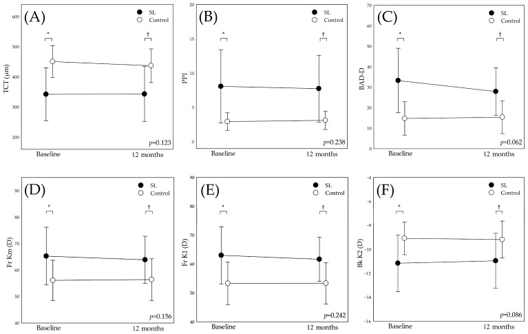
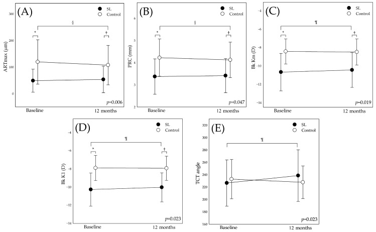
Background and Objectives: Scleral lenses are widely used for visual rehabilitation in keratoconus patients, but their long-term effects on corneal tomography remain unclear. This study aims to evaluate the impact of 12-month scleral lens wear on corneal tomography in keratoconus patients through a case-controlled design. Materials and Methods: This retrospective study included 220 keratoconus patients, of whom 10 eyes were treated with SoClear (Brighten Optix Corporation, Taipei, Taiwan) mini-scleral lenses for over one year (SL group). A control group of 14 eyes was matched using Mahalanobis distance matching based on anterior maximum keratometry (Kmax) and age. Both groups were evaluated at baseline and 12 months. Corneal tomography was assessed using the Pentacam HR (Oculus, Wetzlar, Germany), analyzing parameters such as anterior and posterior corneal curvature, thinnest corneal thickness (TCT), and higher-order aberrations. Generalized estimating equations (GEEs) were employed to assess the time-by-treatment effect between the two groups. Results: The SL group included 10 eyes from eight patients (seven males, one female; mean age 30.40 ± 6.52 years), while the control group included 14 eyes from 11 patients (three males, wight females; mean age 27.43 ± 8.11 years). Best corrected visual acuity with spectacles improved significantly with scleral lenses (p = 0.011) and remained stable (p = 0.044) at 12 months. Significant interaction effects were found in Ambrósio relational thickness (p = 0.006), posterior radius curvature (p = 0.047), posterior mean keratometry (p = 0.019), posterior flat keratometry (p = 0.023), and thinnest corneal thickness angle (p = 0.023); the SL group demonstrated less progression in these parameters compared to the control group. Conclusions: This case-controlled study highlights the 12-month impact of scleral lenses on keratoconus, showing improved visual acuity compared to spectacles, stabilized posterior corneal curvature, and maintained corneal thickness. Further prospective studies with larger cohorts are needed to assess scleral lens effect on keratoconus progression.
期刊介绍:
The journal’s main focus is on reviews as well as clinical and experimental investigations. The journal aims to advance knowledge related to problems in medicine in developing countries as well as developed economies, to disseminate research on global health, and to promote and foster prevention and treatment of diseases worldwide. MEDICINA publications cater to clinicians, diagnosticians and researchers, and serve as a forum to discuss the current status of health-related matters and their impact on a global and local scale.
 求助内容:
求助内容: 应助结果提醒方式:


