{"title":"冠状动脉钙化:类型、形态和分布。","authors":"Michelle C Morris, Rolf P Kreutz","doi":"10.15420/icr.2024.03","DOIUrl":null,"url":null,"abstract":"<p><p>The development and progression of coronary calcification is of growing interest with the emergence of new imaging modalities and calcium modifying technologies that can facilitate optimal results during complex percutaneous coronary intervention (PCI). Coronary atherosclerotic disease typically begins within the intima with pathological intimal thickening and microcalcifications (>0.5 μm and <15 μm). These microcalcifications can coalesce into larger areas of calcification, including sheet calcium, which is typically seen in fibrocalcific plaque, nodular calcification and calcified nodules. Calcified nodules typically protrude into the vessel lumen. Erosive calcified nodules lack the coverage of protective anti-aggregatory endothelium and frequently show adherence of intraluminal thrombus. Greater calcification within coronary plaque does not correlate with an increased risk of acute coronary syndrome, however, coronary calcium can lead to challenges with stent delivery and full stent expansion during PCI. An understanding of plaque morphology, distribution of calcium, degree of calcification and underlying shape will enable interventional cardiologists to appropriately interpret intravascular ultrasound and optical coherence tomography imaging findings and optimise results during complex PCI.</p>","PeriodicalId":38586,"journal":{"name":"Interventional Cardiology Review","volume":"20 ","pages":"e13"},"PeriodicalIF":2.8000,"publicationDate":"2025-04-07","publicationTypes":"Journal Article","fieldsOfStudy":null,"isOpenAccess":false,"openAccessPdf":"https://www.ncbi.nlm.nih.gov/pmc/articles/PMC12042292/pdf/","citationCount":"0","resultStr":"{\"title\":\"Coronary Calcification: Types, Morphology and Distribution.\",\"authors\":\"Michelle C Morris, Rolf P Kreutz\",\"doi\":\"10.15420/icr.2024.03\",\"DOIUrl\":null,\"url\":null,\"abstract\":\"<p><p>The development and progression of coronary calcification is of growing interest with the emergence of new imaging modalities and calcium modifying technologies that can facilitate optimal results during complex percutaneous coronary intervention (PCI). Coronary atherosclerotic disease typically begins within the intima with pathological intimal thickening and microcalcifications (>0.5 μm and <15 μm). These microcalcifications can coalesce into larger areas of calcification, including sheet calcium, which is typically seen in fibrocalcific plaque, nodular calcification and calcified nodules. Calcified nodules typically protrude into the vessel lumen. Erosive calcified nodules lack the coverage of protective anti-aggregatory endothelium and frequently show adherence of intraluminal thrombus. Greater calcification within coronary plaque does not correlate with an increased risk of acute coronary syndrome, however, coronary calcium can lead to challenges with stent delivery and full stent expansion during PCI. An understanding of plaque morphology, distribution of calcium, degree of calcification and underlying shape will enable interventional cardiologists to appropriately interpret intravascular ultrasound and optical coherence tomography imaging findings and optimise results during complex PCI.</p>\",\"PeriodicalId\":38586,\"journal\":{\"name\":\"Interventional Cardiology Review\",\"volume\":\"20 \",\"pages\":\"e13\"},\"PeriodicalIF\":2.8000,\"publicationDate\":\"2025-04-07\",\"publicationTypes\":\"Journal Article\",\"fieldsOfStudy\":null,\"isOpenAccess\":false,\"openAccessPdf\":\"https://www.ncbi.nlm.nih.gov/pmc/articles/PMC12042292/pdf/\",\"citationCount\":\"0\",\"resultStr\":null,\"platform\":\"Semanticscholar\",\"paperid\":null,\"PeriodicalName\":\"Interventional Cardiology Review\",\"FirstCategoryId\":\"1085\",\"ListUrlMain\":\"https://doi.org/10.15420/icr.2024.03\",\"RegionNum\":0,\"RegionCategory\":null,\"ArticlePicture\":[],\"TitleCN\":null,\"AbstractTextCN\":null,\"PMCID\":null,\"EPubDate\":\"2025/1/1 0:00:00\",\"PubModel\":\"eCollection\",\"JCR\":\"0\",\"JCRName\":\"PHILOSOPHY\",\"Score\":null,\"Total\":0}","platform":"Semanticscholar","paperid":null,"PeriodicalName":"Interventional Cardiology Review","FirstCategoryId":"1085","ListUrlMain":"https://doi.org/10.15420/icr.2024.03","RegionNum":0,"RegionCategory":null,"ArticlePicture":[],"TitleCN":null,"AbstractTextCN":null,"PMCID":null,"EPubDate":"2025/1/1 0:00:00","PubModel":"eCollection","JCR":"0","JCRName":"PHILOSOPHY","Score":null,"Total":0}
Coronary Calcification: Types, Morphology and Distribution.
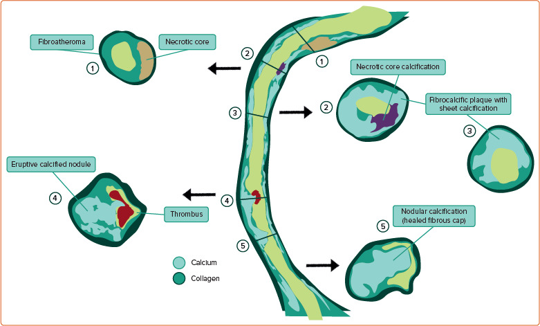
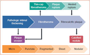
The development and progression of coronary calcification is of growing interest with the emergence of new imaging modalities and calcium modifying technologies that can facilitate optimal results during complex percutaneous coronary intervention (PCI). Coronary atherosclerotic disease typically begins within the intima with pathological intimal thickening and microcalcifications (>0.5 μm and <15 μm). These microcalcifications can coalesce into larger areas of calcification, including sheet calcium, which is typically seen in fibrocalcific plaque, nodular calcification and calcified nodules. Calcified nodules typically protrude into the vessel lumen. Erosive calcified nodules lack the coverage of protective anti-aggregatory endothelium and frequently show adherence of intraluminal thrombus. Greater calcification within coronary plaque does not correlate with an increased risk of acute coronary syndrome, however, coronary calcium can lead to challenges with stent delivery and full stent expansion during PCI. An understanding of plaque morphology, distribution of calcium, degree of calcification and underlying shape will enable interventional cardiologists to appropriately interpret intravascular ultrasound and optical coherence tomography imaging findings and optimise results during complex PCI.

 求助内容:
求助内容: 应助结果提醒方式:


