{"title":"异位胰腺在FAPI PET/CT上伪装成大网膜结节。","authors":"Shrikant Vasantrao Solav, Shailendra Vasant Savale, Hemant Bhagwan Raundale, Vijaykumar Revansidha Keskar","doi":"10.4103/ijnm.ijnm_140_23","DOIUrl":null,"url":null,"abstract":"<p><p>Chylous ascites, attributed to various etiologies including malignancy, tuberculosis, ruptured lymphatics, and congenital lymphatic disorders, manifests as abdominal distension. Our patient presented with this condition, and an elevated CA 125 prompted further investigation. Flourine-18 fluorodeoxyglucose positron emission tomography/computed tomography (PET-CT) revealed a metabolically inactive omental nodule, while gallium 68 fibroblast activation protein inhibitor (Ga-68-FAPI) PET-CT demonstrated uptake in the same nodule and low-grade uptake in bilateral adnexae. Colloid liver scan ruled out chronic liver disease. Surprisingly, lymphoscintigraphy showed no lymphatic leak. Histological examination of the omental nodule confirmed heterotopic pancreas (HP) in the small bowel mesentery, with normal adnexae. This case report illuminates the diagnostic challenges entailed in HP and signifies a pioneering instance in the literature where evidence of HP was identified for the first time on Ga-68-FAPI PET-CT during the investigative process.</p>","PeriodicalId":45830,"journal":{"name":"Indian Journal of Nuclear Medicine","volume":"39 6","pages":"441-444"},"PeriodicalIF":0.5000,"publicationDate":"2024-11-01","publicationTypes":"Journal Article","fieldsOfStudy":null,"isOpenAccess":false,"openAccessPdf":"https://www.ncbi.nlm.nih.gov/pmc/articles/PMC12020968/pdf/","citationCount":"0","resultStr":"{\"title\":\"Heterotopic Pancreas Masquerading as Omental Nodule on FAPI PET/CT.\",\"authors\":\"Shrikant Vasantrao Solav, Shailendra Vasant Savale, Hemant Bhagwan Raundale, Vijaykumar Revansidha Keskar\",\"doi\":\"10.4103/ijnm.ijnm_140_23\",\"DOIUrl\":null,\"url\":null,\"abstract\":\"<p><p>Chylous ascites, attributed to various etiologies including malignancy, tuberculosis, ruptured lymphatics, and congenital lymphatic disorders, manifests as abdominal distension. Our patient presented with this condition, and an elevated CA 125 prompted further investigation. Flourine-18 fluorodeoxyglucose positron emission tomography/computed tomography (PET-CT) revealed a metabolically inactive omental nodule, while gallium 68 fibroblast activation protein inhibitor (Ga-68-FAPI) PET-CT demonstrated uptake in the same nodule and low-grade uptake in bilateral adnexae. Colloid liver scan ruled out chronic liver disease. Surprisingly, lymphoscintigraphy showed no lymphatic leak. Histological examination of the omental nodule confirmed heterotopic pancreas (HP) in the small bowel mesentery, with normal adnexae. This case report illuminates the diagnostic challenges entailed in HP and signifies a pioneering instance in the literature where evidence of HP was identified for the first time on Ga-68-FAPI PET-CT during the investigative process.</p>\",\"PeriodicalId\":45830,\"journal\":{\"name\":\"Indian Journal of Nuclear Medicine\",\"volume\":\"39 6\",\"pages\":\"441-444\"},\"PeriodicalIF\":0.5000,\"publicationDate\":\"2024-11-01\",\"publicationTypes\":\"Journal Article\",\"fieldsOfStudy\":null,\"isOpenAccess\":false,\"openAccessPdf\":\"https://www.ncbi.nlm.nih.gov/pmc/articles/PMC12020968/pdf/\",\"citationCount\":\"0\",\"resultStr\":null,\"platform\":\"Semanticscholar\",\"paperid\":null,\"PeriodicalName\":\"Indian Journal of Nuclear Medicine\",\"FirstCategoryId\":\"1085\",\"ListUrlMain\":\"https://doi.org/10.4103/ijnm.ijnm_140_23\",\"RegionNum\":0,\"RegionCategory\":null,\"ArticlePicture\":[],\"TitleCN\":null,\"AbstractTextCN\":null,\"PMCID\":null,\"EPubDate\":\"2025/3/20 0:00:00\",\"PubModel\":\"Epub\",\"JCR\":\"Q4\",\"JCRName\":\"RADIOLOGY, NUCLEAR MEDICINE & MEDICAL IMAGING\",\"Score\":null,\"Total\":0}","platform":"Semanticscholar","paperid":null,"PeriodicalName":"Indian Journal of Nuclear Medicine","FirstCategoryId":"1085","ListUrlMain":"https://doi.org/10.4103/ijnm.ijnm_140_23","RegionNum":0,"RegionCategory":null,"ArticlePicture":[],"TitleCN":null,"AbstractTextCN":null,"PMCID":null,"EPubDate":"2025/3/20 0:00:00","PubModel":"Epub","JCR":"Q4","JCRName":"RADIOLOGY, NUCLEAR MEDICINE & MEDICAL IMAGING","Score":null,"Total":0}
Heterotopic Pancreas Masquerading as Omental Nodule on FAPI PET/CT.
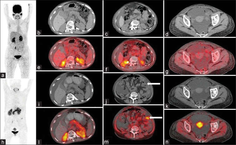
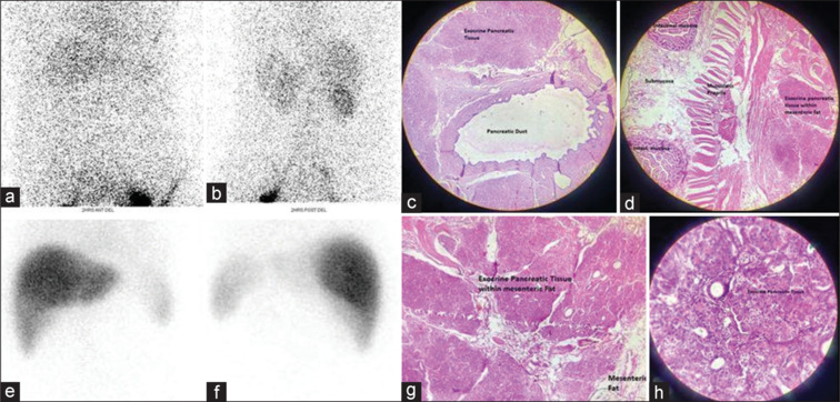
Chylous ascites, attributed to various etiologies including malignancy, tuberculosis, ruptured lymphatics, and congenital lymphatic disorders, manifests as abdominal distension. Our patient presented with this condition, and an elevated CA 125 prompted further investigation. Flourine-18 fluorodeoxyglucose positron emission tomography/computed tomography (PET-CT) revealed a metabolically inactive omental nodule, while gallium 68 fibroblast activation protein inhibitor (Ga-68-FAPI) PET-CT demonstrated uptake in the same nodule and low-grade uptake in bilateral adnexae. Colloid liver scan ruled out chronic liver disease. Surprisingly, lymphoscintigraphy showed no lymphatic leak. Histological examination of the omental nodule confirmed heterotopic pancreas (HP) in the small bowel mesentery, with normal adnexae. This case report illuminates the diagnostic challenges entailed in HP and signifies a pioneering instance in the literature where evidence of HP was identified for the first time on Ga-68-FAPI PET-CT during the investigative process.

 求助内容:
求助内容: 应助结果提醒方式:


