Arthur Ho, Lisa Feng, Jos J Rozema, David A Atchison
{"title":"眼底成像中多节段晶状体的视网膜阴影。","authors":"Arthur Ho, Lisa Feng, Jos J Rozema, David A Atchison","doi":"10.1111/opo.13524","DOIUrl":null,"url":null,"abstract":"<p><strong>Purpose: </strong>To provide a more sophisticated explanation of the optics involved when retinal 'shadows' are seen in scanning laser ophthalmoscopic images during the wear of multisegment and diffusion optic spectacle lenses.</p><p><strong>Methods: </strong>Images were recorded with a system that uses a scanning broad line fundus imaging principle in participants with undilated pupils wearing a multisegment spectacle lens. The live infra-red preview display of the system was also acquired during image recording. Ray-tracing and image simulations were performed, assuming a Maxwellian illumination system in which a source was refracted first through a lens and then through a model of a multisegment spectacle lens focused onto the pupil of an eye model and hence to the retina. A detector surface was positioned slightly in front of the retina to record the irradiation distribution. The light source was varied from 0.1 μm to 1.8 mm in diameter to investigate the effect of light source size on retinal irradiation distribution.</p><p><strong>Results: </strong>The retinal shadow pattern was visible on the live infra-red preview display, as reported previously. However, the recorded images of the retina do not exhibit the same shadow pattern. The simulations predict that the circular shadows corresponding to lenslet positions become progressively less discernible with increasing light source size.</p><p><strong>Conclusion: </strong>An explanation is provided for the shadows on retinal images due to multisegment lenses, which may be observable under certain illumination conditions.</p>","PeriodicalId":19522,"journal":{"name":"Ophthalmic and Physiological Optics","volume":" ","pages":"1098-1103"},"PeriodicalIF":2.4000,"publicationDate":"2025-07-01","publicationTypes":"Journal Article","fieldsOfStudy":null,"isOpenAccess":false,"openAccessPdf":"https://www.ncbi.nlm.nih.gov/pmc/articles/PMC12153018/pdf/","citationCount":"0","resultStr":"{\"title\":\"Retinal shadows of multisegment lenses in fundus imaging.\",\"authors\":\"Arthur Ho, Lisa Feng, Jos J Rozema, David A Atchison\",\"doi\":\"10.1111/opo.13524\",\"DOIUrl\":null,\"url\":null,\"abstract\":\"<p><strong>Purpose: </strong>To provide a more sophisticated explanation of the optics involved when retinal 'shadows' are seen in scanning laser ophthalmoscopic images during the wear of multisegment and diffusion optic spectacle lenses.</p><p><strong>Methods: </strong>Images were recorded with a system that uses a scanning broad line fundus imaging principle in participants with undilated pupils wearing a multisegment spectacle lens. The live infra-red preview display of the system was also acquired during image recording. Ray-tracing and image simulations were performed, assuming a Maxwellian illumination system in which a source was refracted first through a lens and then through a model of a multisegment spectacle lens focused onto the pupil of an eye model and hence to the retina. A detector surface was positioned slightly in front of the retina to record the irradiation distribution. The light source was varied from 0.1 μm to 1.8 mm in diameter to investigate the effect of light source size on retinal irradiation distribution.</p><p><strong>Results: </strong>The retinal shadow pattern was visible on the live infra-red preview display, as reported previously. However, the recorded images of the retina do not exhibit the same shadow pattern. The simulations predict that the circular shadows corresponding to lenslet positions become progressively less discernible with increasing light source size.</p><p><strong>Conclusion: </strong>An explanation is provided for the shadows on retinal images due to multisegment lenses, which may be observable under certain illumination conditions.</p>\",\"PeriodicalId\":19522,\"journal\":{\"name\":\"Ophthalmic and Physiological Optics\",\"volume\":\" \",\"pages\":\"1098-1103\"},\"PeriodicalIF\":2.4000,\"publicationDate\":\"2025-07-01\",\"publicationTypes\":\"Journal Article\",\"fieldsOfStudy\":null,\"isOpenAccess\":false,\"openAccessPdf\":\"https://www.ncbi.nlm.nih.gov/pmc/articles/PMC12153018/pdf/\",\"citationCount\":\"0\",\"resultStr\":null,\"platform\":\"Semanticscholar\",\"paperid\":null,\"PeriodicalName\":\"Ophthalmic and Physiological Optics\",\"FirstCategoryId\":\"3\",\"ListUrlMain\":\"https://doi.org/10.1111/opo.13524\",\"RegionNum\":3,\"RegionCategory\":\"医学\",\"ArticlePicture\":[],\"TitleCN\":null,\"AbstractTextCN\":null,\"PMCID\":null,\"EPubDate\":\"2025/5/14 0:00:00\",\"PubModel\":\"Epub\",\"JCR\":\"Q1\",\"JCRName\":\"OPHTHALMOLOGY\",\"Score\":null,\"Total\":0}","platform":"Semanticscholar","paperid":null,"PeriodicalName":"Ophthalmic and Physiological Optics","FirstCategoryId":"3","ListUrlMain":"https://doi.org/10.1111/opo.13524","RegionNum":3,"RegionCategory":"医学","ArticlePicture":[],"TitleCN":null,"AbstractTextCN":null,"PMCID":null,"EPubDate":"2025/5/14 0:00:00","PubModel":"Epub","JCR":"Q1","JCRName":"OPHTHALMOLOGY","Score":null,"Total":0}
Retinal shadows of multisegment lenses in fundus imaging.
Purpose: To provide a more sophisticated explanation of the optics involved when retinal 'shadows' are seen in scanning laser ophthalmoscopic images during the wear of multisegment and diffusion optic spectacle lenses.
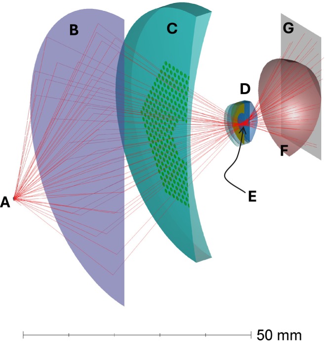
Methods: Images were recorded with a system that uses a scanning broad line fundus imaging principle in participants with undilated pupils wearing a multisegment spectacle lens. The live infra-red preview display of the system was also acquired during image recording. Ray-tracing and image simulations were performed, assuming a Maxwellian illumination system in which a source was refracted first through a lens and then through a model of a multisegment spectacle lens focused onto the pupil of an eye model and hence to the retina. A detector surface was positioned slightly in front of the retina to record the irradiation distribution. The light source was varied from 0.1 μm to 1.8 mm in diameter to investigate the effect of light source size on retinal irradiation distribution.
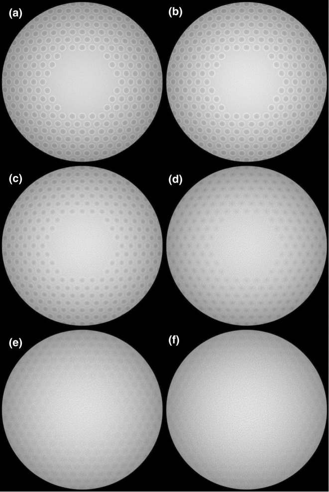
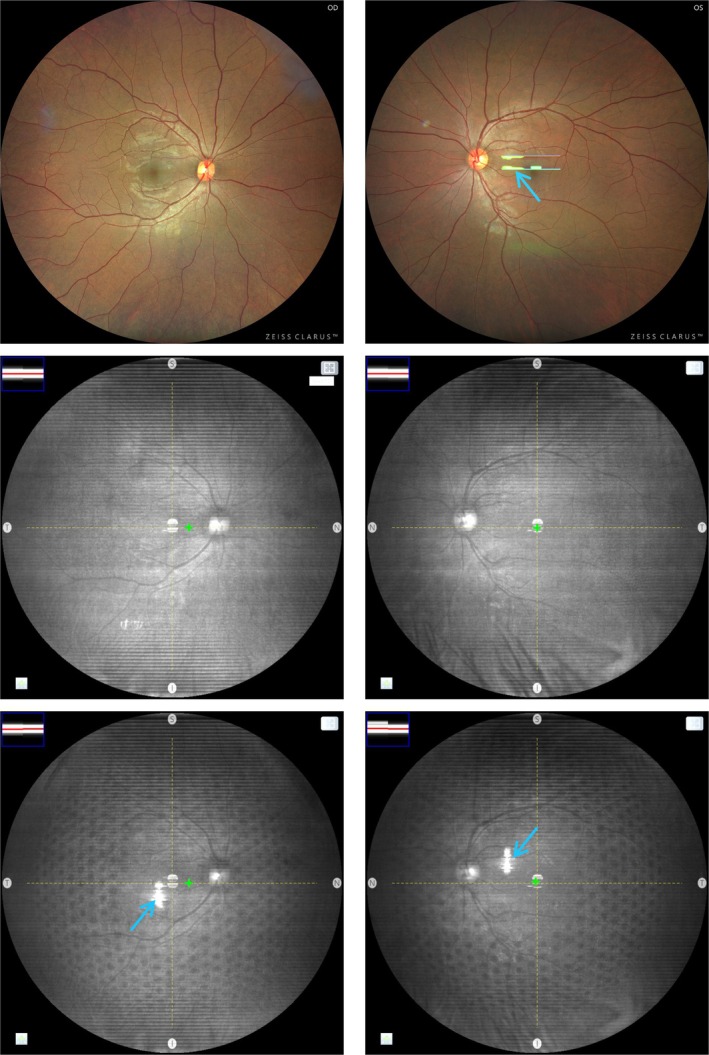
Results: The retinal shadow pattern was visible on the live infra-red preview display, as reported previously. However, the recorded images of the retina do not exhibit the same shadow pattern. The simulations predict that the circular shadows corresponding to lenslet positions become progressively less discernible with increasing light source size.
Conclusion: An explanation is provided for the shadows on retinal images due to multisegment lenses, which may be observable under certain illumination conditions.
期刊介绍:
Ophthalmic & Physiological Optics, first published in 1925, is a leading international interdisciplinary journal that addresses basic and applied questions pertinent to contemporary research in vision science and optometry.
OPO publishes original research papers, technical notes, reviews and letters and will interest researchers, educators and clinicians concerned with the development, use and restoration of vision.
 求助内容:
求助内容: 应助结果提醒方式:


