Yanzhuo Li, Sijie Li, Yan Lei, Lianlian Liu, Bin Song
{"title":"磁共振成像增强模式与生长模式的结合评价肝细胞癌微血管浸润的严重程度。","authors":"Yanzhuo Li, Sijie Li, Yan Lei, Lianlian Liu, Bin Song","doi":"10.2478/raon-2025-0021","DOIUrl":null,"url":null,"abstract":"<p><strong>Background: </strong>Microvascular invasion (MVI), particularly its severity, correlates with prognosis in hepatocellular carcinoma (HCC), however, it remains uncertain which imaging traits are associated with MVI grades. Predicting MVI status precisely pre-surgery assists clinicians in making optimal treatment decisions.</p><p><strong>Patients and methods: </strong>213 HCC patients with surgically confirmed were assigned into three groups based on the severity of MVI (M0, M1, and M2). Clinical and imaging features were compared between each group. Univariate and multivariate analyses were used to identify the significant variables associated with MVI severity. Subsequently, nomograms were constructed to estimate MVI and its M2 grade by crucial factors. Nomograms were assessed for accuracy, clinical value, and efficacy using the area under the curve (AUC), calibration curve, and decision curve analysis (DCA).</p><p><strong>Results: </strong>Four factors associated with MVI (P < 0.05) were related, including non-solitary growth types, no/mini enhanced mode, peritumoral enhancement on arterial phase, and peritumoral hypointensity on hepatobiliary phase. Only the ratio of the maximum and minimum tumor diameter (Max/Min-R), confluent multinodule growth type, and non-washin/washout enhanced modes of those MVI-positive patients showed a strong correlation with M2 grade. The areas under the receiver operating characteristic (ROC) curves were 0.885 (95% confidence intervals [CI]: 0.833-0.937) in identifying MVI and 0.805 (95% CI: 0.703-0.908) in predicting its M2 grade, respectively. The nomograms demonstrated a high goodness-of-fit and clinical benefits in DCA and calibration curve.</p><p><strong>Conclusions: </strong>Enhancement modes and tumor growth patterns of preoperative MRI were independent risk factors of MVI severity, which were valuable for facilitating individualized decision-making.</p>","PeriodicalId":21034,"journal":{"name":"Radiology and Oncology","volume":" ","pages":"183-192"},"PeriodicalIF":2.2000,"publicationDate":"2025-04-11","publicationTypes":"Journal Article","fieldsOfStudy":null,"isOpenAccess":false,"openAccessPdf":"https://www.ncbi.nlm.nih.gov/pmc/articles/PMC12182925/pdf/","citationCount":"0","resultStr":"{\"title\":\"Evaluating the severity of microvascular invasion in hepatocellular carcinoma, by probing the combination of enhancement modes and growth patterns through magnetic resonance imaging.\",\"authors\":\"Yanzhuo Li, Sijie Li, Yan Lei, Lianlian Liu, Bin Song\",\"doi\":\"10.2478/raon-2025-0021\",\"DOIUrl\":null,\"url\":null,\"abstract\":\"<p><strong>Background: </strong>Microvascular invasion (MVI), particularly its severity, correlates with prognosis in hepatocellular carcinoma (HCC), however, it remains uncertain which imaging traits are associated with MVI grades. Predicting MVI status precisely pre-surgery assists clinicians in making optimal treatment decisions.</p><p><strong>Patients and methods: </strong>213 HCC patients with surgically confirmed were assigned into three groups based on the severity of MVI (M0, M1, and M2). Clinical and imaging features were compared between each group. Univariate and multivariate analyses were used to identify the significant variables associated with MVI severity. Subsequently, nomograms were constructed to estimate MVI and its M2 grade by crucial factors. Nomograms were assessed for accuracy, clinical value, and efficacy using the area under the curve (AUC), calibration curve, and decision curve analysis (DCA).</p><p><strong>Results: </strong>Four factors associated with MVI (P < 0.05) were related, including non-solitary growth types, no/mini enhanced mode, peritumoral enhancement on arterial phase, and peritumoral hypointensity on hepatobiliary phase. Only the ratio of the maximum and minimum tumor diameter (Max/Min-R), confluent multinodule growth type, and non-washin/washout enhanced modes of those MVI-positive patients showed a strong correlation with M2 grade. The areas under the receiver operating characteristic (ROC) curves were 0.885 (95% confidence intervals [CI]: 0.833-0.937) in identifying MVI and 0.805 (95% CI: 0.703-0.908) in predicting its M2 grade, respectively. The nomograms demonstrated a high goodness-of-fit and clinical benefits in DCA and calibration curve.</p><p><strong>Conclusions: </strong>Enhancement modes and tumor growth patterns of preoperative MRI were independent risk factors of MVI severity, which were valuable for facilitating individualized decision-making.</p>\",\"PeriodicalId\":21034,\"journal\":{\"name\":\"Radiology and Oncology\",\"volume\":\" \",\"pages\":\"183-192\"},\"PeriodicalIF\":2.2000,\"publicationDate\":\"2025-04-11\",\"publicationTypes\":\"Journal Article\",\"fieldsOfStudy\":null,\"isOpenAccess\":false,\"openAccessPdf\":\"https://www.ncbi.nlm.nih.gov/pmc/articles/PMC12182925/pdf/\",\"citationCount\":\"0\",\"resultStr\":null,\"platform\":\"Semanticscholar\",\"paperid\":null,\"PeriodicalName\":\"Radiology and Oncology\",\"FirstCategoryId\":\"3\",\"ListUrlMain\":\"https://doi.org/10.2478/raon-2025-0021\",\"RegionNum\":4,\"RegionCategory\":\"医学\",\"ArticlePicture\":[],\"TitleCN\":null,\"AbstractTextCN\":null,\"PMCID\":null,\"EPubDate\":\"2025/6/1 0:00:00\",\"PubModel\":\"eCollection\",\"JCR\":\"Q3\",\"JCRName\":\"ONCOLOGY\",\"Score\":null,\"Total\":0}","platform":"Semanticscholar","paperid":null,"PeriodicalName":"Radiology and Oncology","FirstCategoryId":"3","ListUrlMain":"https://doi.org/10.2478/raon-2025-0021","RegionNum":4,"RegionCategory":"医学","ArticlePicture":[],"TitleCN":null,"AbstractTextCN":null,"PMCID":null,"EPubDate":"2025/6/1 0:00:00","PubModel":"eCollection","JCR":"Q3","JCRName":"ONCOLOGY","Score":null,"Total":0}
Evaluating the severity of microvascular invasion in hepatocellular carcinoma, by probing the combination of enhancement modes and growth patterns through magnetic resonance imaging.
Background: Microvascular invasion (MVI), particularly its severity, correlates with prognosis in hepatocellular carcinoma (HCC), however, it remains uncertain which imaging traits are associated with MVI grades. Predicting MVI status precisely pre-surgery assists clinicians in making optimal treatment decisions.
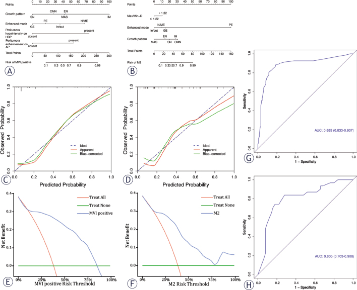
Patients and methods: 213 HCC patients with surgically confirmed were assigned into three groups based on the severity of MVI (M0, M1, and M2). Clinical and imaging features were compared between each group. Univariate and multivariate analyses were used to identify the significant variables associated with MVI severity. Subsequently, nomograms were constructed to estimate MVI and its M2 grade by crucial factors. Nomograms were assessed for accuracy, clinical value, and efficacy using the area under the curve (AUC), calibration curve, and decision curve analysis (DCA).
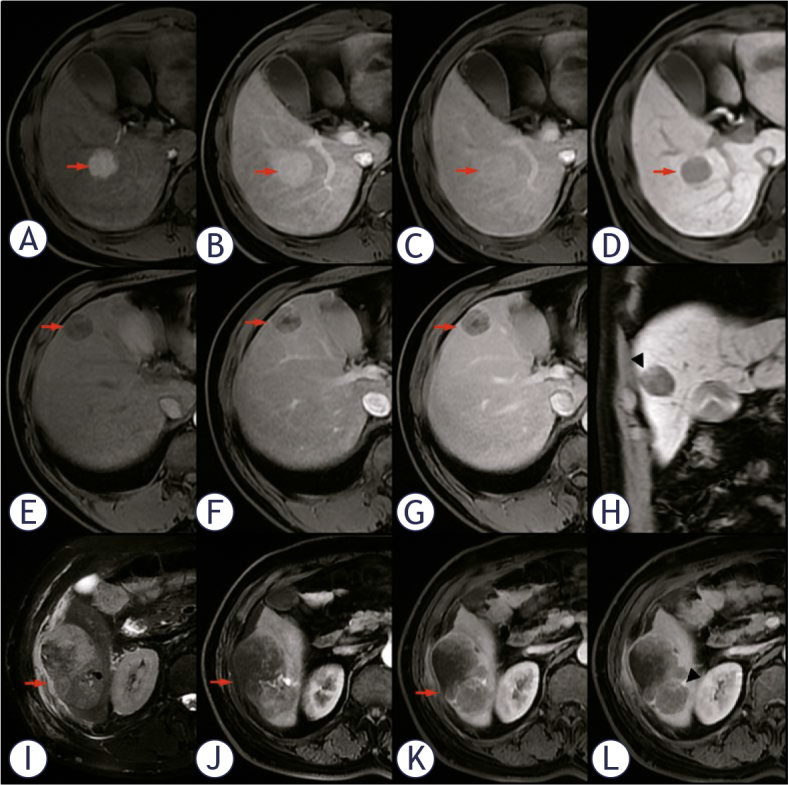
Results: Four factors associated with MVI (P < 0.05) were related, including non-solitary growth types, no/mini enhanced mode, peritumoral enhancement on arterial phase, and peritumoral hypointensity on hepatobiliary phase. Only the ratio of the maximum and minimum tumor diameter (Max/Min-R), confluent multinodule growth type, and non-washin/washout enhanced modes of those MVI-positive patients showed a strong correlation with M2 grade. The areas under the receiver operating characteristic (ROC) curves were 0.885 (95% confidence intervals [CI]: 0.833-0.937) in identifying MVI and 0.805 (95% CI: 0.703-0.908) in predicting its M2 grade, respectively. The nomograms demonstrated a high goodness-of-fit and clinical benefits in DCA and calibration curve.
Conclusions: Enhancement modes and tumor growth patterns of preoperative MRI were independent risk factors of MVI severity, which were valuable for facilitating individualized decision-making.
期刊介绍:
Radiology and Oncology is a multidisciplinary journal devoted to the publishing original and high quality scientific papers and review articles, pertinent to diagnostic and interventional radiology, computerized tomography, magnetic resonance, ultrasound, nuclear medicine, radiotherapy, clinical and experimental oncology, radiobiology, medical physics and radiation protection. Therefore, the scope of the journal is to cover beside radiology the diagnostic and therapeutic aspects in oncology, which distinguishes it from other journals in the field.
 求助内容:
求助内容: 应助结果提醒方式:


