Beth Whittington, Viswan Thiagarajah, Evangelos Tzolos, Jakub Kaczynski, Caelan Taggart, Alex Vesey, Damini Dey, Rachael O Forsythe, Andrew Tambyraja, Edwin J R van Beek, Marc R Dweck, David E Newby, Michelle C Williams
{"title":"定量颈动脉斑块和血管周围脂肪组织衰减的计算机断层扫描。","authors":"Beth Whittington, Viswan Thiagarajah, Evangelos Tzolos, Jakub Kaczynski, Caelan Taggart, Alex Vesey, Damini Dey, Rachael O Forsythe, Andrew Tambyraja, Edwin J R van Beek, Marc R Dweck, David E Newby, Michelle C Williams","doi":"10.1093/ehjimp/qyaf040","DOIUrl":null,"url":null,"abstract":"<p><strong>Aims: </strong>Quantitative assessment of carotid artery plaque on computed tomography (CT) may identify high-risk phenotypes associated with culprit lesions and subsequent ischaemic stroke or transient ischaemic attack.</p><p><strong>Methods and results: </strong>Carotid CT angiography was performed in 48 patients with acute ischaemic stroke or transient ischaemic attack within 21 days. Quantitative plaque assessment was performed in the proximal 6 cm of the internal and external carotid artery, distal 6 cm of the common carotid artery, and residual common carotid artery. Semi-automated quantification included assessment of non-calcified, calcified, low-attenuation, and total plaque, area and diameter stenosis, and peri-vascular adipose tissue attenuation. In 48 patients (mean age 71 ± 11 years, 67% male), 96 vessels were assessed with 30 (31%) identified as culprit vessels. Culprit internal carotid arteries had greater area [83 (65, 94) vs. 64 (55, 77)%] and diameter [56 (39, 74) vs. 32 (21, 48)%] stenosis and more non-calcified [563 (413, 965) vs. 428 (283 649) mm<sup>3</sup>, <i>P</i> = 0.04], low-attenuation [33.7 (6.9, 72.4) vs. 16.3 (3.35, 54.3) mm<sup>3</sup>, <i>P</i> = 0.01], and total [699 (455, 1057) vs. 492 (311, 809), <i>P</i> = 0.04] plaque. There was no difference in calcified plaque or peri-vascular adipose tissue attenuation between culprit and non-culprit internal carotid arteries. There were no differences in quantitative plaque or peri-vascular adipose tissue attenuation in the external carotid artery or common carotid artery.</p><p><strong>Conclusion: </strong>Carotid atherosclerotic plaque characteristics are the principal features associated with culprit plaques with little or no demonstrable relationship with calcified plaque or increased peri-vascular adipose tissue attenuation.</p>","PeriodicalId":94317,"journal":{"name":"European heart journal. Imaging methods and practice","volume":"3 1","pages":"qyaf040"},"PeriodicalIF":0.0000,"publicationDate":"2025-04-08","publicationTypes":"Journal Article","fieldsOfStudy":null,"isOpenAccess":false,"openAccessPdf":"https://www.ncbi.nlm.nih.gov/pmc/articles/PMC12023745/pdf/","citationCount":"0","resultStr":"{\"title\":\"Quantification of carotid artery plaque and peri-vascular adipose tissue attenuation on computed tomography.\",\"authors\":\"Beth Whittington, Viswan Thiagarajah, Evangelos Tzolos, Jakub Kaczynski, Caelan Taggart, Alex Vesey, Damini Dey, Rachael O Forsythe, Andrew Tambyraja, Edwin J R van Beek, Marc R Dweck, David E Newby, Michelle C Williams\",\"doi\":\"10.1093/ehjimp/qyaf040\",\"DOIUrl\":null,\"url\":null,\"abstract\":\"<p><strong>Aims: </strong>Quantitative assessment of carotid artery plaque on computed tomography (CT) may identify high-risk phenotypes associated with culprit lesions and subsequent ischaemic stroke or transient ischaemic attack.</p><p><strong>Methods and results: </strong>Carotid CT angiography was performed in 48 patients with acute ischaemic stroke or transient ischaemic attack within 21 days. Quantitative plaque assessment was performed in the proximal 6 cm of the internal and external carotid artery, distal 6 cm of the common carotid artery, and residual common carotid artery. Semi-automated quantification included assessment of non-calcified, calcified, low-attenuation, and total plaque, area and diameter stenosis, and peri-vascular adipose tissue attenuation. In 48 patients (mean age 71 ± 11 years, 67% male), 96 vessels were assessed with 30 (31%) identified as culprit vessels. Culprit internal carotid arteries had greater area [83 (65, 94) vs. 64 (55, 77)%] and diameter [56 (39, 74) vs. 32 (21, 48)%] stenosis and more non-calcified [563 (413, 965) vs. 428 (283 649) mm<sup>3</sup>, <i>P</i> = 0.04], low-attenuation [33.7 (6.9, 72.4) vs. 16.3 (3.35, 54.3) mm<sup>3</sup>, <i>P</i> = 0.01], and total [699 (455, 1057) vs. 492 (311, 809), <i>P</i> = 0.04] plaque. There was no difference in calcified plaque or peri-vascular adipose tissue attenuation between culprit and non-culprit internal carotid arteries. There were no differences in quantitative plaque or peri-vascular adipose tissue attenuation in the external carotid artery or common carotid artery.</p><p><strong>Conclusion: </strong>Carotid atherosclerotic plaque characteristics are the principal features associated with culprit plaques with little or no demonstrable relationship with calcified plaque or increased peri-vascular adipose tissue attenuation.</p>\",\"PeriodicalId\":94317,\"journal\":{\"name\":\"European heart journal. Imaging methods and practice\",\"volume\":\"3 1\",\"pages\":\"qyaf040\"},\"PeriodicalIF\":0.0000,\"publicationDate\":\"2025-04-08\",\"publicationTypes\":\"Journal Article\",\"fieldsOfStudy\":null,\"isOpenAccess\":false,\"openAccessPdf\":\"https://www.ncbi.nlm.nih.gov/pmc/articles/PMC12023745/pdf/\",\"citationCount\":\"0\",\"resultStr\":null,\"platform\":\"Semanticscholar\",\"paperid\":null,\"PeriodicalName\":\"European heart journal. Imaging methods and practice\",\"FirstCategoryId\":\"1085\",\"ListUrlMain\":\"https://doi.org/10.1093/ehjimp/qyaf040\",\"RegionNum\":0,\"RegionCategory\":null,\"ArticlePicture\":[],\"TitleCN\":null,\"AbstractTextCN\":null,\"PMCID\":null,\"EPubDate\":\"2025/1/1 0:00:00\",\"PubModel\":\"eCollection\",\"JCR\":\"\",\"JCRName\":\"\",\"Score\":null,\"Total\":0}","platform":"Semanticscholar","paperid":null,"PeriodicalName":"European heart journal. Imaging methods and practice","FirstCategoryId":"1085","ListUrlMain":"https://doi.org/10.1093/ehjimp/qyaf040","RegionNum":0,"RegionCategory":null,"ArticlePicture":[],"TitleCN":null,"AbstractTextCN":null,"PMCID":null,"EPubDate":"2025/1/1 0:00:00","PubModel":"eCollection","JCR":"","JCRName":"","Score":null,"Total":0}
引用次数: 0
摘要
目的:通过计算机断层扫描(CT)对颈动脉斑块进行定量评估,可以识别与罪魁祸首病变和随后的缺血性卒中或短暂性缺血性发作相关的高危表型。方法与结果:对48例急性缺血性脑卒中或短暂性缺血性发作患者在21天内行颈动脉CT血管造影。定量评估颈内、外动脉近端6cm、颈总动脉远端6cm及颈总动脉残余斑块。半自动化量化包括评估非钙化、钙化、低衰减和总斑块、面积和直径狭窄以及血管周围脂肪组织衰减。48例患者(平均年龄71±11岁,男性67%),96条血管被评估,其中30条(31%)被确定为罪魁祸首血管。罪魁祸首颈内动脉狭窄面积增大[83(65,94)比64(55,77)%],直径增大[56(39,74)比32(21,48)%],非钙化斑块增多[563(413,965)比428 (283,649)mm3, P = 0.04],低衰减斑块[33.7(6.9,72.4)比16.3 (3.35,54.3)mm3, P = 0.01],斑块总数[699(455,1057)比492 (311,809),P = 0.04]。罪魁祸首和非罪魁祸首颈内动脉的钙化斑块和血管周围脂肪组织衰减没有差异。颈外动脉和颈总动脉斑块数量和血管周围脂肪组织衰减没有差异。结论:颈动脉粥样硬化斑块特征是罪魁祸首斑块的主要特征,与钙化斑块或血管周围脂肪组织衰减很少或没有明显的关系。
Quantification of carotid artery plaque and peri-vascular adipose tissue attenuation on computed tomography.
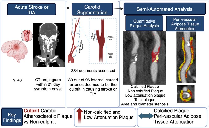
Aims: Quantitative assessment of carotid artery plaque on computed tomography (CT) may identify high-risk phenotypes associated with culprit lesions and subsequent ischaemic stroke or transient ischaemic attack.
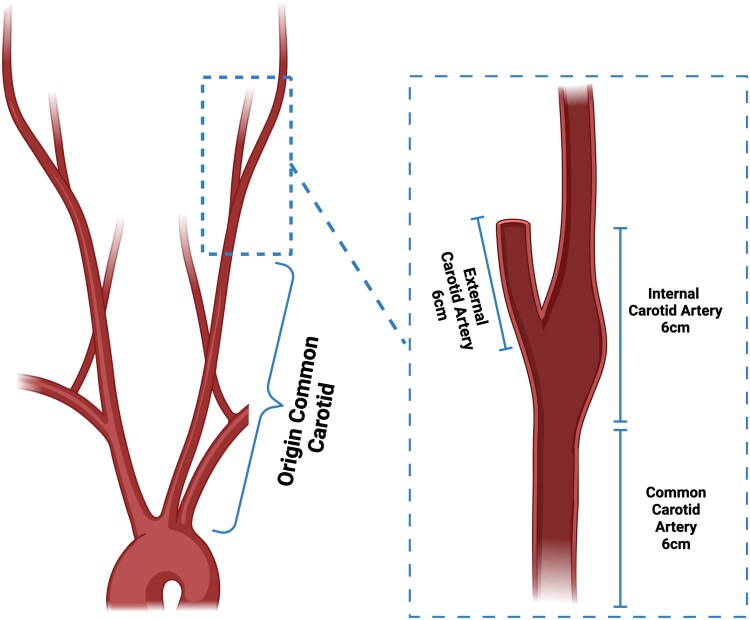
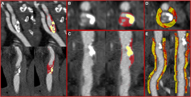
Methods and results: Carotid CT angiography was performed in 48 patients with acute ischaemic stroke or transient ischaemic attack within 21 days. Quantitative plaque assessment was performed in the proximal 6 cm of the internal and external carotid artery, distal 6 cm of the common carotid artery, and residual common carotid artery. Semi-automated quantification included assessment of non-calcified, calcified, low-attenuation, and total plaque, area and diameter stenosis, and peri-vascular adipose tissue attenuation. In 48 patients (mean age 71 ± 11 years, 67% male), 96 vessels were assessed with 30 (31%) identified as culprit vessels. Culprit internal carotid arteries had greater area [83 (65, 94) vs. 64 (55, 77)%] and diameter [56 (39, 74) vs. 32 (21, 48)%] stenosis and more non-calcified [563 (413, 965) vs. 428 (283 649) mm3, P = 0.04], low-attenuation [33.7 (6.9, 72.4) vs. 16.3 (3.35, 54.3) mm3, P = 0.01], and total [699 (455, 1057) vs. 492 (311, 809), P = 0.04] plaque. There was no difference in calcified plaque or peri-vascular adipose tissue attenuation between culprit and non-culprit internal carotid arteries. There were no differences in quantitative plaque or peri-vascular adipose tissue attenuation in the external carotid artery or common carotid artery.
Conclusion: Carotid atherosclerotic plaque characteristics are the principal features associated with culprit plaques with little or no demonstrable relationship with calcified plaque or increased peri-vascular adipose tissue attenuation.

 求助内容:
求助内容: 应助结果提醒方式:


