Amberpreet K Khangura, Shally Gupta, Anubha Gulati, Manjula Mehta
{"title":"钙荧光白和吖啶橙染色检测口腔潜在恶性疾病和口腔鳞状细胞癌真菌成分的比较评价——回顾性研究。","authors":"Amberpreet K Khangura, Shally Gupta, Anubha Gulati, Manjula Mehta","doi":"10.4103/jomfp.jomfp_117_24","DOIUrl":null,"url":null,"abstract":"<p><strong>Background: </strong>Candida is an opportunistic fungal pathogen that frequently colonizes the oral mucosa. Depending on multiple etiological factors, Candida can transform from a harmless commensal into a pathogenic organism, leading to the development of oral potentially malignant disorders (OPMDs) and oral cancer. Although various laboratory diagnostic methods for Candida have been developed, there remains a need for more rapid and sensitive diagnostic aids for infections associated with Candida. The present study aimed to compare the efficacy of Calcofluor white and acridine orange fluorescent stains and evaluate the most efficacious stain.</p><p><strong>Materials and methods: </strong>Two fluorescent stains, calcofluor white and acridine orange, were used to identify Candida elements in formalin-fixed, paraffin-embedded tissue sections of oral potentially malignant disorders (n = 16), including leukoplakia without dysplasia, mild dysplasia, moderate dysplasia, severe dysplasia, oral lichen planus, and oral sub mucous fibrosis as well as oral squamous cell carcinoma (n = 16), encompassing well-differentiated, moderately differentiated, and poorly differentiated grades. All stained slides were examined using a fluorescence microscope equipped with a blue filter at 20× and 40× magnifications. The comparison between the two stains was conducted based on the expression of fungal elements or the grade of Candida within the given sections, staining quality, and cost-effectiveness.</p><p><strong>Results: </strong>The results of the present study confirmed that both stains produced similar outcomes in terms of the expression of fungal elements within the sections of oral potentially malignant disorders and oral squamous cell carcinoma. Using both stains, all cases were positive (n = 32), with no negative cases reported. Grade I and II Candida was identified in the sections of oral potentially malignant disorders, whereas Grade III and IV Candida were observed within the sections of oral squamous cell carcinoma and sever dysplasia. Calcofluor white stain demonstrated higher efficiency in terms of staining quality and cost-effectiveness.</p><p><strong>Conclusion: </strong>Calcofluor white stain exhibited better expression of Candida elements in cases of oral potentially malignant disorders and oral cancer in terms of staining efficacy and is also more cost-effective.</p>","PeriodicalId":38846,"journal":{"name":"Journal of Oral and Maxillofacial Pathology","volume":"29 1","pages":"98-103"},"PeriodicalIF":0.0000,"publicationDate":"2025-01-01","publicationTypes":"Journal Article","fieldsOfStudy":null,"isOpenAccess":false,"openAccessPdf":"https://www.ncbi.nlm.nih.gov/pmc/articles/PMC12002577/pdf/","citationCount":"0","resultStr":"{\"title\":\"Comparative evaluation of staining efficacy of calcofluor white and acridine orange in detecting fungal elements in oral potentially malignant disorders and oral squamous cell carcinoma - A retrospective study.\",\"authors\":\"Amberpreet K Khangura, Shally Gupta, Anubha Gulati, Manjula Mehta\",\"doi\":\"10.4103/jomfp.jomfp_117_24\",\"DOIUrl\":null,\"url\":null,\"abstract\":\"<p><strong>Background: </strong>Candida is an opportunistic fungal pathogen that frequently colonizes the oral mucosa. Depending on multiple etiological factors, Candida can transform from a harmless commensal into a pathogenic organism, leading to the development of oral potentially malignant disorders (OPMDs) and oral cancer. Although various laboratory diagnostic methods for Candida have been developed, there remains a need for more rapid and sensitive diagnostic aids for infections associated with Candida. The present study aimed to compare the efficacy of Calcofluor white and acridine orange fluorescent stains and evaluate the most efficacious stain.</p><p><strong>Materials and methods: </strong>Two fluorescent stains, calcofluor white and acridine orange, were used to identify Candida elements in formalin-fixed, paraffin-embedded tissue sections of oral potentially malignant disorders (n = 16), including leukoplakia without dysplasia, mild dysplasia, moderate dysplasia, severe dysplasia, oral lichen planus, and oral sub mucous fibrosis as well as oral squamous cell carcinoma (n = 16), encompassing well-differentiated, moderately differentiated, and poorly differentiated grades. All stained slides were examined using a fluorescence microscope equipped with a blue filter at 20× and 40× magnifications. The comparison between the two stains was conducted based on the expression of fungal elements or the grade of Candida within the given sections, staining quality, and cost-effectiveness.</p><p><strong>Results: </strong>The results of the present study confirmed that both stains produced similar outcomes in terms of the expression of fungal elements within the sections of oral potentially malignant disorders and oral squamous cell carcinoma. Using both stains, all cases were positive (n = 32), with no negative cases reported. Grade I and II Candida was identified in the sections of oral potentially malignant disorders, whereas Grade III and IV Candida were observed within the sections of oral squamous cell carcinoma and sever dysplasia. Calcofluor white stain demonstrated higher efficiency in terms of staining quality and cost-effectiveness.</p><p><strong>Conclusion: </strong>Calcofluor white stain exhibited better expression of Candida elements in cases of oral potentially malignant disorders and oral cancer in terms of staining efficacy and is also more cost-effective.</p>\",\"PeriodicalId\":38846,\"journal\":{\"name\":\"Journal of Oral and Maxillofacial Pathology\",\"volume\":\"29 1\",\"pages\":\"98-103\"},\"PeriodicalIF\":0.0000,\"publicationDate\":\"2025-01-01\",\"publicationTypes\":\"Journal Article\",\"fieldsOfStudy\":null,\"isOpenAccess\":false,\"openAccessPdf\":\"https://www.ncbi.nlm.nih.gov/pmc/articles/PMC12002577/pdf/\",\"citationCount\":\"0\",\"resultStr\":null,\"platform\":\"Semanticscholar\",\"paperid\":null,\"PeriodicalName\":\"Journal of Oral and Maxillofacial Pathology\",\"FirstCategoryId\":\"1085\",\"ListUrlMain\":\"https://doi.org/10.4103/jomfp.jomfp_117_24\",\"RegionNum\":0,\"RegionCategory\":null,\"ArticlePicture\":[],\"TitleCN\":null,\"AbstractTextCN\":null,\"PMCID\":null,\"EPubDate\":\"2025/3/28 0:00:00\",\"PubModel\":\"Epub\",\"JCR\":\"Q3\",\"JCRName\":\"Medicine\",\"Score\":null,\"Total\":0}","platform":"Semanticscholar","paperid":null,"PeriodicalName":"Journal of Oral and Maxillofacial Pathology","FirstCategoryId":"1085","ListUrlMain":"https://doi.org/10.4103/jomfp.jomfp_117_24","RegionNum":0,"RegionCategory":null,"ArticlePicture":[],"TitleCN":null,"AbstractTextCN":null,"PMCID":null,"EPubDate":"2025/3/28 0:00:00","PubModel":"Epub","JCR":"Q3","JCRName":"Medicine","Score":null,"Total":0}
Comparative evaluation of staining efficacy of calcofluor white and acridine orange in detecting fungal elements in oral potentially malignant disorders and oral squamous cell carcinoma - A retrospective study.
Background: Candida is an opportunistic fungal pathogen that frequently colonizes the oral mucosa. Depending on multiple etiological factors, Candida can transform from a harmless commensal into a pathogenic organism, leading to the development of oral potentially malignant disorders (OPMDs) and oral cancer. Although various laboratory diagnostic methods for Candida have been developed, there remains a need for more rapid and sensitive diagnostic aids for infections associated with Candida. The present study aimed to compare the efficacy of Calcofluor white and acridine orange fluorescent stains and evaluate the most efficacious stain.
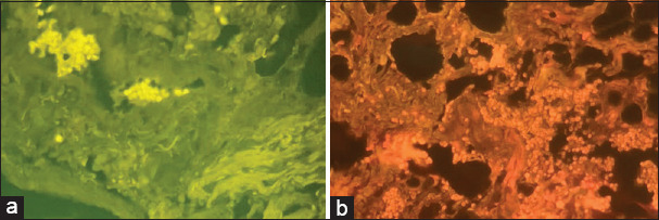
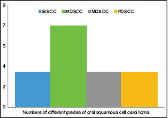
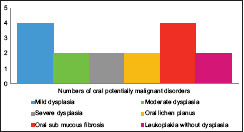
Materials and methods: Two fluorescent stains, calcofluor white and acridine orange, were used to identify Candida elements in formalin-fixed, paraffin-embedded tissue sections of oral potentially malignant disorders (n = 16), including leukoplakia without dysplasia, mild dysplasia, moderate dysplasia, severe dysplasia, oral lichen planus, and oral sub mucous fibrosis as well as oral squamous cell carcinoma (n = 16), encompassing well-differentiated, moderately differentiated, and poorly differentiated grades. All stained slides were examined using a fluorescence microscope equipped with a blue filter at 20× and 40× magnifications. The comparison between the two stains was conducted based on the expression of fungal elements or the grade of Candida within the given sections, staining quality, and cost-effectiveness.
Results: The results of the present study confirmed that both stains produced similar outcomes in terms of the expression of fungal elements within the sections of oral potentially malignant disorders and oral squamous cell carcinoma. Using both stains, all cases were positive (n = 32), with no negative cases reported. Grade I and II Candida was identified in the sections of oral potentially malignant disorders, whereas Grade III and IV Candida were observed within the sections of oral squamous cell carcinoma and sever dysplasia. Calcofluor white stain demonstrated higher efficiency in terms of staining quality and cost-effectiveness.
Conclusion: Calcofluor white stain exhibited better expression of Candida elements in cases of oral potentially malignant disorders and oral cancer in terms of staining efficacy and is also more cost-effective.
期刊介绍:
The journal of Oral and Maxillofacial Pathology [ISSN:print-(0973-029X, online-1998-393X)] is a tri-annual journal published on behalf of “The Indian Association of Oral and Maxillofacial Pathologists” (IAOMP). The publication of JOMFP was started in the year 1993. The journal publishes papers on a wide spectrum of topics associated with the scope of Oral and Maxillofacial Pathology, also, ensuring scientific merit and quality. It is a comprehensive reading material for the professionals who want to upgrade their diagnostic skills in Oral Diseases; allows exposure to newer topics and methods of research in the Oral-facial Tissues and Pathology. New features allow an open minded thinking and approach to various pathologies. It also encourages authors to showcase quality work done by them and to compile relevant cases which are diagnostically challenging. The Journal takes pride in maintaining the quality of articles and photomicrographs.

 求助内容:
求助内容: 应助结果提醒方式:


