Shubhanshi Trivedi, Olivia J Cheng, Ben J Brintz, Richelle C Charles, Daniel T Leung
{"title":"肠沙门氏菌血清型伤寒Ty21a口服疫苗接种者的粘膜相关不变性T (MAIT)细胞反应","authors":"Shubhanshi Trivedi, Olivia J Cheng, Ben J Brintz, Richelle C Charles, Daniel T Leung","doi":"10.1093/oxfimm/iqaf002","DOIUrl":null,"url":null,"abstract":"<p><p>Mucosal-associated invariant T (MAIT) cells are unconventional innate-like T cells abundant in human mucosal tissues and are associated with protective responses to microbial infections. MAIT cells have the capacity for rapid effector functions, including the secretion of cytokines and cytotoxic molecules. In this study, we examined the longitudinal circulating MAIT cell response to the live attenuated oral vaccine Ty21a (Ty21a) against <i>Salmonella enterica</i> serovar Typhi (<i>S</i>. Typhi). We enrolled healthy adults who received a course of oral live-attenuated <i>S.</i> Typhi strain Ty21a vaccine and assessed peripheral blood MAIT cell longitudinal responses pre-vaccination, and at seven days and one-month post-vaccination, using flow cytometry, cell migration, and tetramer decay assays. We showed that following vaccination, circulating MAIT cells were lower in frequency, but were more activated, and had higher levels of gut-homing marker integrin α4β7 and chemokine receptors CCR9 and CCR6, suggesting the potential of MAIT cells to migrate to mucosal sites. We found no significant differences in MAIT cell functionality, cytotoxicity and T-cell receptor avidity, except in TNF expression, which was higher post-vaccination. We show that MAIT cell immune responses are modulated post-vaccination against <i>S.</i> Typhi. This study contributes to our understanding of MAIT cells' potential role in oral vaccination against bacterial mucosal pathogens.</p>","PeriodicalId":74384,"journal":{"name":"Oxford open immunology","volume":"6 1","pages":"iqaf002"},"PeriodicalIF":0.0000,"publicationDate":"2025-03-25","publicationTypes":"Journal Article","fieldsOfStudy":null,"isOpenAccess":false,"openAccessPdf":"https://www.ncbi.nlm.nih.gov/pmc/articles/PMC11993846/pdf/","citationCount":"0","resultStr":"{\"title\":\"Mucosal-associated invariant T (MAIT) cell responses in <i>Salmonella enterica</i> serovar Typhi strain Ty21a oral vaccine recipients.\",\"authors\":\"Shubhanshi Trivedi, Olivia J Cheng, Ben J Brintz, Richelle C Charles, Daniel T Leung\",\"doi\":\"10.1093/oxfimm/iqaf002\",\"DOIUrl\":null,\"url\":null,\"abstract\":\"<p><p>Mucosal-associated invariant T (MAIT) cells are unconventional innate-like T cells abundant in human mucosal tissues and are associated with protective responses to microbial infections. MAIT cells have the capacity for rapid effector functions, including the secretion of cytokines and cytotoxic molecules. In this study, we examined the longitudinal circulating MAIT cell response to the live attenuated oral vaccine Ty21a (Ty21a) against <i>Salmonella enterica</i> serovar Typhi (<i>S</i>. Typhi). We enrolled healthy adults who received a course of oral live-attenuated <i>S.</i> Typhi strain Ty21a vaccine and assessed peripheral blood MAIT cell longitudinal responses pre-vaccination, and at seven days and one-month post-vaccination, using flow cytometry, cell migration, and tetramer decay assays. We showed that following vaccination, circulating MAIT cells were lower in frequency, but were more activated, and had higher levels of gut-homing marker integrin α4β7 and chemokine receptors CCR9 and CCR6, suggesting the potential of MAIT cells to migrate to mucosal sites. We found no significant differences in MAIT cell functionality, cytotoxicity and T-cell receptor avidity, except in TNF expression, which was higher post-vaccination. We show that MAIT cell immune responses are modulated post-vaccination against <i>S.</i> Typhi. This study contributes to our understanding of MAIT cells' potential role in oral vaccination against bacterial mucosal pathogens.</p>\",\"PeriodicalId\":74384,\"journal\":{\"name\":\"Oxford open immunology\",\"volume\":\"6 1\",\"pages\":\"iqaf002\"},\"PeriodicalIF\":0.0000,\"publicationDate\":\"2025-03-25\",\"publicationTypes\":\"Journal Article\",\"fieldsOfStudy\":null,\"isOpenAccess\":false,\"openAccessPdf\":\"https://www.ncbi.nlm.nih.gov/pmc/articles/PMC11993846/pdf/\",\"citationCount\":\"0\",\"resultStr\":null,\"platform\":\"Semanticscholar\",\"paperid\":null,\"PeriodicalName\":\"Oxford open immunology\",\"FirstCategoryId\":\"1085\",\"ListUrlMain\":\"https://doi.org/10.1093/oxfimm/iqaf002\",\"RegionNum\":0,\"RegionCategory\":null,\"ArticlePicture\":[],\"TitleCN\":null,\"AbstractTextCN\":null,\"PMCID\":null,\"EPubDate\":\"2025/1/1 0:00:00\",\"PubModel\":\"eCollection\",\"JCR\":\"\",\"JCRName\":\"\",\"Score\":null,\"Total\":0}","platform":"Semanticscholar","paperid":null,"PeriodicalName":"Oxford open immunology","FirstCategoryId":"1085","ListUrlMain":"https://doi.org/10.1093/oxfimm/iqaf002","RegionNum":0,"RegionCategory":null,"ArticlePicture":[],"TitleCN":null,"AbstractTextCN":null,"PMCID":null,"EPubDate":"2025/1/1 0:00:00","PubModel":"eCollection","JCR":"","JCRName":"","Score":null,"Total":0}
Mucosal-associated invariant T (MAIT) cell responses in Salmonella enterica serovar Typhi strain Ty21a oral vaccine recipients.
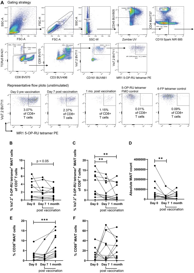
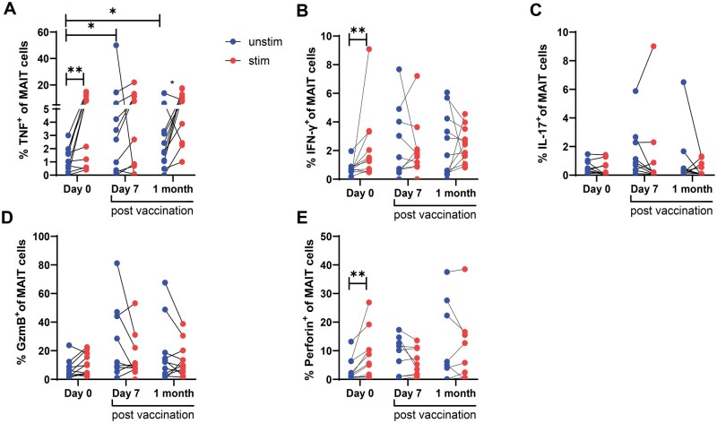
Mucosal-associated invariant T (MAIT) cells are unconventional innate-like T cells abundant in human mucosal tissues and are associated with protective responses to microbial infections. MAIT cells have the capacity for rapid effector functions, including the secretion of cytokines and cytotoxic molecules. In this study, we examined the longitudinal circulating MAIT cell response to the live attenuated oral vaccine Ty21a (Ty21a) against Salmonella enterica serovar Typhi (S. Typhi). We enrolled healthy adults who received a course of oral live-attenuated S. Typhi strain Ty21a vaccine and assessed peripheral blood MAIT cell longitudinal responses pre-vaccination, and at seven days and one-month post-vaccination, using flow cytometry, cell migration, and tetramer decay assays. We showed that following vaccination, circulating MAIT cells were lower in frequency, but were more activated, and had higher levels of gut-homing marker integrin α4β7 and chemokine receptors CCR9 and CCR6, suggesting the potential of MAIT cells to migrate to mucosal sites. We found no significant differences in MAIT cell functionality, cytotoxicity and T-cell receptor avidity, except in TNF expression, which was higher post-vaccination. We show that MAIT cell immune responses are modulated post-vaccination against S. Typhi. This study contributes to our understanding of MAIT cells' potential role in oral vaccination against bacterial mucosal pathogens.

 求助内容:
求助内容: 应助结果提醒方式:


