{"title":"系统性红斑狼疮患者淋巴结中的朗格汉斯细胞浸润。","authors":"Jinyi Qian, Lei Li, Jing Lv, Yingjie Jiang, Qianchen Ma, Haoyu Pan, Xiaohan Wei, Zhixia Yang, Shuyi Yu, Yuying Fan, Jialin Teng, Chengde Yang, Aifei Zhang, Yue Yang, Hui Shi","doi":"10.1136/lupus-2024-001474","DOIUrl":null,"url":null,"abstract":"<p><strong>Objective: </strong>This study aimed to characterise the clinical features and treatment regimens of patients with lupus who have lymphadenopathy (LAP), as well as to investigate the presence and potential implications of Langerhans cells (LCs) infiltration in lymph nodes.</p><p><strong>Methods: </strong>A case-control study was conducted to identify the clinical characteristics of newly diagnosed, treatment-naïve patients with lupus who have LAP. Lymph node biopsies were performed, and LC infiltration was assessed using immunohistochemical staining for S100, CD1a and Langerin.</p><p><strong>Results: </strong>A total of 59 patients with SLE who have LAP (SLE-LAP) were enrolled, with 81 patients with SLE without LAP serving as controls. The SLE-LAP group exhibited significantly higher frequencies of fever (64.4% vs 35.8%, p<0.001), anaemia (71.2% vs 42.0%, p<0.001), serous effusion (27.1% vs 11.1%, p=0.015), myositis (10.2% vs 1.2%, p=0.045) and elevated CRP levels (44.1% vs 22.2%, p=0.006). Moreover, autoantibodies, including anti-Smith (37.3% vs 16.0%, p=0.004), anticardiolipin IgG (27.1% vs 11.1%, p=0.015), IgM (42.4% vs 9.9%, p<0.001) and IgA (8.5% vs 0.0%, p=0.027), were more frequently detected in the LAP group. LC infiltration was confirmed in 29 of the 59 lymph node biopsies (49.2%). Immunohistochemical analysis revealed a scattered (58.6%) or focal (41.4%) distribution of LCs. Patients with LC infiltration predominantly presented with fever (72.4%), anaemia (64.3%), skin rashes (62.1%) and arthritis (62.1%). However, no significant differences in clinical manifestations were observed between LC-positive and LC-negative patients.</p><p><strong>Conclusion: </strong>LC infiltration in the lymph nodes of patients with SLE is relatively common and should be carefully evaluated to prevent misdiagnosis. The role of LCs in the autoimmune response and pathogenesis of SLE warrants further investigation.</p>","PeriodicalId":18126,"journal":{"name":"Lupus Science & Medicine","volume":"12 1","pages":""},"PeriodicalIF":3.5000,"publicationDate":"2025-04-14","publicationTypes":"Journal Article","fieldsOfStudy":null,"isOpenAccess":false,"openAccessPdf":"https://www.ncbi.nlm.nih.gov/pmc/articles/PMC11997825/pdf/","citationCount":"0","resultStr":"{\"title\":\"Langerhans cells infiltration in lymph nodes of patients with systemic lupus erythematosus.\",\"authors\":\"Jinyi Qian, Lei Li, Jing Lv, Yingjie Jiang, Qianchen Ma, Haoyu Pan, Xiaohan Wei, Zhixia Yang, Shuyi Yu, Yuying Fan, Jialin Teng, Chengde Yang, Aifei Zhang, Yue Yang, Hui Shi\",\"doi\":\"10.1136/lupus-2024-001474\",\"DOIUrl\":null,\"url\":null,\"abstract\":\"<p><strong>Objective: </strong>This study aimed to characterise the clinical features and treatment regimens of patients with lupus who have lymphadenopathy (LAP), as well as to investigate the presence and potential implications of Langerhans cells (LCs) infiltration in lymph nodes.</p><p><strong>Methods: </strong>A case-control study was conducted to identify the clinical characteristics of newly diagnosed, treatment-naïve patients with lupus who have LAP. Lymph node biopsies were performed, and LC infiltration was assessed using immunohistochemical staining for S100, CD1a and Langerin.</p><p><strong>Results: </strong>A total of 59 patients with SLE who have LAP (SLE-LAP) were enrolled, with 81 patients with SLE without LAP serving as controls. The SLE-LAP group exhibited significantly higher frequencies of fever (64.4% vs 35.8%, p<0.001), anaemia (71.2% vs 42.0%, p<0.001), serous effusion (27.1% vs 11.1%, p=0.015), myositis (10.2% vs 1.2%, p=0.045) and elevated CRP levels (44.1% vs 22.2%, p=0.006). Moreover, autoantibodies, including anti-Smith (37.3% vs 16.0%, p=0.004), anticardiolipin IgG (27.1% vs 11.1%, p=0.015), IgM (42.4% vs 9.9%, p<0.001) and IgA (8.5% vs 0.0%, p=0.027), were more frequently detected in the LAP group. LC infiltration was confirmed in 29 of the 59 lymph node biopsies (49.2%). Immunohistochemical analysis revealed a scattered (58.6%) or focal (41.4%) distribution of LCs. Patients with LC infiltration predominantly presented with fever (72.4%), anaemia (64.3%), skin rashes (62.1%) and arthritis (62.1%). However, no significant differences in clinical manifestations were observed between LC-positive and LC-negative patients.</p><p><strong>Conclusion: </strong>LC infiltration in the lymph nodes of patients with SLE is relatively common and should be carefully evaluated to prevent misdiagnosis. The role of LCs in the autoimmune response and pathogenesis of SLE warrants further investigation.</p>\",\"PeriodicalId\":18126,\"journal\":{\"name\":\"Lupus Science & Medicine\",\"volume\":\"12 1\",\"pages\":\"\"},\"PeriodicalIF\":3.5000,\"publicationDate\":\"2025-04-14\",\"publicationTypes\":\"Journal Article\",\"fieldsOfStudy\":null,\"isOpenAccess\":false,\"openAccessPdf\":\"https://www.ncbi.nlm.nih.gov/pmc/articles/PMC11997825/pdf/\",\"citationCount\":\"0\",\"resultStr\":null,\"platform\":\"Semanticscholar\",\"paperid\":null,\"PeriodicalName\":\"Lupus Science & Medicine\",\"FirstCategoryId\":\"3\",\"ListUrlMain\":\"https://doi.org/10.1136/lupus-2024-001474\",\"RegionNum\":2,\"RegionCategory\":\"医学\",\"ArticlePicture\":[],\"TitleCN\":null,\"AbstractTextCN\":null,\"PMCID\":null,\"EPubDate\":\"\",\"PubModel\":\"\",\"JCR\":\"Q1\",\"JCRName\":\"RHEUMATOLOGY\",\"Score\":null,\"Total\":0}","platform":"Semanticscholar","paperid":null,"PeriodicalName":"Lupus Science & Medicine","FirstCategoryId":"3","ListUrlMain":"https://doi.org/10.1136/lupus-2024-001474","RegionNum":2,"RegionCategory":"医学","ArticlePicture":[],"TitleCN":null,"AbstractTextCN":null,"PMCID":null,"EPubDate":"","PubModel":"","JCR":"Q1","JCRName":"RHEUMATOLOGY","Score":null,"Total":0}
引用次数: 0
摘要
目的:本研究旨在探讨红斑狼疮伴淋巴结病变(LAP)患者的临床特征和治疗方案,以及淋巴结中朗格汉斯细胞(LCs)浸润的存在及其潜在意义。方法:通过病例对照研究,确定新诊断的treatment-naïve狼疮LAP患者的临床特征。行淋巴结活检,采用免疫组化S100、CD1a和Langerin染色评估LC浸润情况。结果:共有59例合并LAP (SLE-LAP)的SLE患者入组,81例未合并LAP的SLE患者作为对照组。SLE- lap组发热发生率明显高于对照组(64.4% vs 35.8%)。结论:SLE患者淋巴结LC浸润较为常见,应仔细评估,防止误诊。LCs在SLE自身免疫反应和发病机制中的作用有待进一步研究。
Langerhans cells infiltration in lymph nodes of patients with systemic lupus erythematosus.
Objective: This study aimed to characterise the clinical features and treatment regimens of patients with lupus who have lymphadenopathy (LAP), as well as to investigate the presence and potential implications of Langerhans cells (LCs) infiltration in lymph nodes.
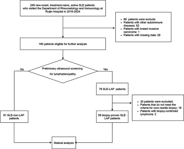
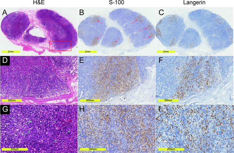
Methods: A case-control study was conducted to identify the clinical characteristics of newly diagnosed, treatment-naïve patients with lupus who have LAP. Lymph node biopsies were performed, and LC infiltration was assessed using immunohistochemical staining for S100, CD1a and Langerin.
Results: A total of 59 patients with SLE who have LAP (SLE-LAP) were enrolled, with 81 patients with SLE without LAP serving as controls. The SLE-LAP group exhibited significantly higher frequencies of fever (64.4% vs 35.8%, p<0.001), anaemia (71.2% vs 42.0%, p<0.001), serous effusion (27.1% vs 11.1%, p=0.015), myositis (10.2% vs 1.2%, p=0.045) and elevated CRP levels (44.1% vs 22.2%, p=0.006). Moreover, autoantibodies, including anti-Smith (37.3% vs 16.0%, p=0.004), anticardiolipin IgG (27.1% vs 11.1%, p=0.015), IgM (42.4% vs 9.9%, p<0.001) and IgA (8.5% vs 0.0%, p=0.027), were more frequently detected in the LAP group. LC infiltration was confirmed in 29 of the 59 lymph node biopsies (49.2%). Immunohistochemical analysis revealed a scattered (58.6%) or focal (41.4%) distribution of LCs. Patients with LC infiltration predominantly presented with fever (72.4%), anaemia (64.3%), skin rashes (62.1%) and arthritis (62.1%). However, no significant differences in clinical manifestations were observed between LC-positive and LC-negative patients.
Conclusion: LC infiltration in the lymph nodes of patients with SLE is relatively common and should be carefully evaluated to prevent misdiagnosis. The role of LCs in the autoimmune response and pathogenesis of SLE warrants further investigation.
期刊介绍:
Lupus Science & Medicine is a global, peer reviewed, open access online journal that provides a central point for publication of basic, clinical, translational, and epidemiological studies of all aspects of lupus and related diseases. It is the first lupus-specific open access journal in the world and was developed in response to the need for a barrier-free forum for publication of groundbreaking studies in lupus. The journal publishes research on lupus from fields including, but not limited to: rheumatology, dermatology, nephrology, immunology, pediatrics, cardiology, hepatology, pulmonology, obstetrics and gynecology, and psychiatry.
 求助内容:
求助内容: 应助结果提醒方式:


