Mohamed Elshibly, Simran Shergill, Kelly Parke, Charley Budgeon, Rachel England, Ciaran Grafton-Clarke, Fathelrahman Elshibly, Peter Kellman, Gerry P McCann, Jayanth R Arnold
{"title":"标准屏气与自由呼吸实时心脏mri -已知或疑似心脏病患者的前瞻性随机比较","authors":"Mohamed Elshibly, Simran Shergill, Kelly Parke, Charley Budgeon, Rachel England, Ciaran Grafton-Clarke, Fathelrahman Elshibly, Peter Kellman, Gerry P McCann, Jayanth R Arnold","doi":"10.1093/ehjimp/qyaf042","DOIUrl":null,"url":null,"abstract":"<p><strong>Aims: </strong>Cardiovascular magnetic resonance (CMR) is established as the reference standard for cardiac volumetric assessment. Despite the accuracy and robustness of steady-state free precession (SSFP) cine imaging, its use may prove challenging in patients with arrhythmia and in those who cannot perform repeated breath holds. An alternative solution may be a free-breathing electrocardiogram (ECG)-triggered, retro-gated, real-time cine sequence. This study sought to compare left ventricular volumetric, wall motion, and thickness assessment with both techniques.</p><p><strong>Methods and results: </strong>Consecutive patients with known or suspected cardiac disease referred for clinical CMR were studied at 3-Tesla. Participants underwent short-axis standard SSFP and real-time cine imaging in a randomized order within the same scan. Between sequence agreement and mean difference were compared for end-diastolic volume (EDV), end-systolic volume (ESV), stroke volume, ejection fraction (EF), left ventricular mass (LVM), maximal wall thickness (MWT), and wall motion score index (WMSi). Two hundred and two patients (mean age 61 ± 14 years, 51% male and 14% irregular rhythm) were studied. All left ventricular indices showed good-excellent agreement between the two methods [intraclass correlation coefficient (95% confidence interval), EDV 0.96 (0.95-0.97), ESV 0.96 (0.94-0.97), EF 0.85 (0.81-0.88), LVM 0.93 (0.91-0.95), MWT 0.80 (0.75-0.85), and WMSi 0.93 (0.91-0.95)].</p><p><strong>Conclusion: </strong>In patients with known or suspected cardiac disease, real-time cine imaging demonstrates good-excellent reproducibility of LV volumetric, wall thickness and resting wall motion assessment when compared with standard SSFP (Trial registration: NCT05221853).</p>","PeriodicalId":94317,"journal":{"name":"European heart journal. Imaging methods and practice","volume":"3 1","pages":"qyaf042"},"PeriodicalIF":0.0000,"publicationDate":"2025-04-25","publicationTypes":"Journal Article","fieldsOfStudy":null,"isOpenAccess":false,"openAccessPdf":"https://www.ncbi.nlm.nih.gov/pmc/articles/PMC12041914/pdf/","citationCount":"0","resultStr":"{\"title\":\"Standard breath-hold versus free-breathing real-time cine cardiac MRI-a prospective randomized comparison in patients with known or suspected cardiac disease.\",\"authors\":\"Mohamed Elshibly, Simran Shergill, Kelly Parke, Charley Budgeon, Rachel England, Ciaran Grafton-Clarke, Fathelrahman Elshibly, Peter Kellman, Gerry P McCann, Jayanth R Arnold\",\"doi\":\"10.1093/ehjimp/qyaf042\",\"DOIUrl\":null,\"url\":null,\"abstract\":\"<p><strong>Aims: </strong>Cardiovascular magnetic resonance (CMR) is established as the reference standard for cardiac volumetric assessment. Despite the accuracy and robustness of steady-state free precession (SSFP) cine imaging, its use may prove challenging in patients with arrhythmia and in those who cannot perform repeated breath holds. An alternative solution may be a free-breathing electrocardiogram (ECG)-triggered, retro-gated, real-time cine sequence. This study sought to compare left ventricular volumetric, wall motion, and thickness assessment with both techniques.</p><p><strong>Methods and results: </strong>Consecutive patients with known or suspected cardiac disease referred for clinical CMR were studied at 3-Tesla. Participants underwent short-axis standard SSFP and real-time cine imaging in a randomized order within the same scan. Between sequence agreement and mean difference were compared for end-diastolic volume (EDV), end-systolic volume (ESV), stroke volume, ejection fraction (EF), left ventricular mass (LVM), maximal wall thickness (MWT), and wall motion score index (WMSi). Two hundred and two patients (mean age 61 ± 14 years, 51% male and 14% irregular rhythm) were studied. All left ventricular indices showed good-excellent agreement between the two methods [intraclass correlation coefficient (95% confidence interval), EDV 0.96 (0.95-0.97), ESV 0.96 (0.94-0.97), EF 0.85 (0.81-0.88), LVM 0.93 (0.91-0.95), MWT 0.80 (0.75-0.85), and WMSi 0.93 (0.91-0.95)].</p><p><strong>Conclusion: </strong>In patients with known or suspected cardiac disease, real-time cine imaging demonstrates good-excellent reproducibility of LV volumetric, wall thickness and resting wall motion assessment when compared with standard SSFP (Trial registration: NCT05221853).</p>\",\"PeriodicalId\":94317,\"journal\":{\"name\":\"European heart journal. Imaging methods and practice\",\"volume\":\"3 1\",\"pages\":\"qyaf042\"},\"PeriodicalIF\":0.0000,\"publicationDate\":\"2025-04-25\",\"publicationTypes\":\"Journal Article\",\"fieldsOfStudy\":null,\"isOpenAccess\":false,\"openAccessPdf\":\"https://www.ncbi.nlm.nih.gov/pmc/articles/PMC12041914/pdf/\",\"citationCount\":\"0\",\"resultStr\":null,\"platform\":\"Semanticscholar\",\"paperid\":null,\"PeriodicalName\":\"European heart journal. Imaging methods and practice\",\"FirstCategoryId\":\"1085\",\"ListUrlMain\":\"https://doi.org/10.1093/ehjimp/qyaf042\",\"RegionNum\":0,\"RegionCategory\":null,\"ArticlePicture\":[],\"TitleCN\":null,\"AbstractTextCN\":null,\"PMCID\":null,\"EPubDate\":\"2025/1/1 0:00:00\",\"PubModel\":\"eCollection\",\"JCR\":\"\",\"JCRName\":\"\",\"Score\":null,\"Total\":0}","platform":"Semanticscholar","paperid":null,"PeriodicalName":"European heart journal. Imaging methods and practice","FirstCategoryId":"1085","ListUrlMain":"https://doi.org/10.1093/ehjimp/qyaf042","RegionNum":0,"RegionCategory":null,"ArticlePicture":[],"TitleCN":null,"AbstractTextCN":null,"PMCID":null,"EPubDate":"2025/1/1 0:00:00","PubModel":"eCollection","JCR":"","JCRName":"","Score":null,"Total":0}
Standard breath-hold versus free-breathing real-time cine cardiac MRI-a prospective randomized comparison in patients with known or suspected cardiac disease.
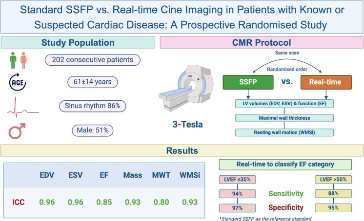
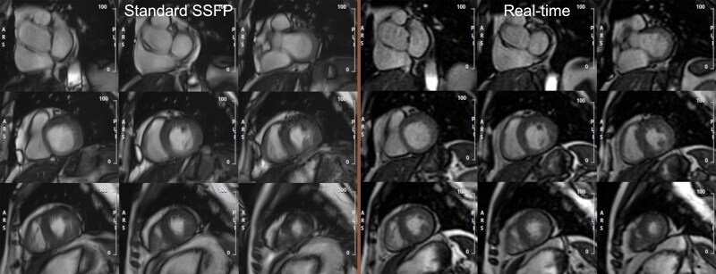
Aims: Cardiovascular magnetic resonance (CMR) is established as the reference standard for cardiac volumetric assessment. Despite the accuracy and robustness of steady-state free precession (SSFP) cine imaging, its use may prove challenging in patients with arrhythmia and in those who cannot perform repeated breath holds. An alternative solution may be a free-breathing electrocardiogram (ECG)-triggered, retro-gated, real-time cine sequence. This study sought to compare left ventricular volumetric, wall motion, and thickness assessment with both techniques.
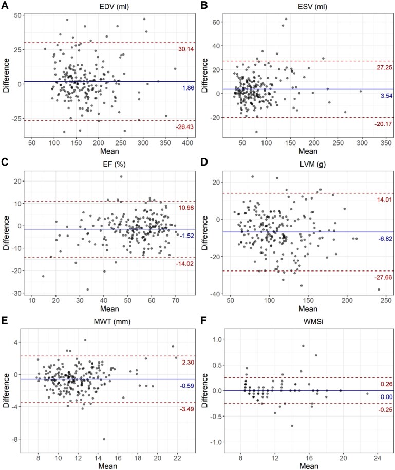
Methods and results: Consecutive patients with known or suspected cardiac disease referred for clinical CMR were studied at 3-Tesla. Participants underwent short-axis standard SSFP and real-time cine imaging in a randomized order within the same scan. Between sequence agreement and mean difference were compared for end-diastolic volume (EDV), end-systolic volume (ESV), stroke volume, ejection fraction (EF), left ventricular mass (LVM), maximal wall thickness (MWT), and wall motion score index (WMSi). Two hundred and two patients (mean age 61 ± 14 years, 51% male and 14% irregular rhythm) were studied. All left ventricular indices showed good-excellent agreement between the two methods [intraclass correlation coefficient (95% confidence interval), EDV 0.96 (0.95-0.97), ESV 0.96 (0.94-0.97), EF 0.85 (0.81-0.88), LVM 0.93 (0.91-0.95), MWT 0.80 (0.75-0.85), and WMSi 0.93 (0.91-0.95)].
Conclusion: In patients with known or suspected cardiac disease, real-time cine imaging demonstrates good-excellent reproducibility of LV volumetric, wall thickness and resting wall motion assessment when compared with standard SSFP (Trial registration: NCT05221853).

 求助内容:
求助内容: 应助结果提醒方式:


