Bruna Bressianini Lima, Rafael Kretzer Carneiro, Brenda Santos Pompeu Miranda, Beatriz Gasser, Luiz Paulo Nogueira Aires, Verônica Maria Teixeira de Castro Terrabuio, Ricardo Andrés Ramirez Uscategui, Antônio Carlos Cunha Lacreta Junior, Danuta Pulz Doiche, Gabriela Castro Lopes Evangelista, Marcus Antônio Rossi Feliciano
{"title":"狗肺部病变的声辐射力脉冲弹性图研究。","authors":"Bruna Bressianini Lima, Rafael Kretzer Carneiro, Brenda Santos Pompeu Miranda, Beatriz Gasser, Luiz Paulo Nogueira Aires, Verônica Maria Teixeira de Castro Terrabuio, Ricardo Andrés Ramirez Uscategui, Antônio Carlos Cunha Lacreta Junior, Danuta Pulz Doiche, Gabriela Castro Lopes Evangelista, Marcus Antônio Rossi Feliciano","doi":"10.1111/vru.70031","DOIUrl":null,"url":null,"abstract":"<p><p>This study aimed to evaluate the use of acoustic radiation force impulse (ARFI) elastography as a diagnostic tool for lung lesions in dogs. Dogs referred to the Radiology Department of the Veterinary Teaching Hospital between 2020 and 2022 for the detection of lung lesions were included in the study. The characteristics of the lung lesions were assessed using radiography as a screening tool for localization, B-mode ultrasound for tissue characterization, and subsequently, both qualitative (elastogram grades 1-3) and quantitative (shear wave velocity-SWV) elastographic evaluations. The lesions were classified based on clinical, ultrasound, radiographic, histopathological, and/or cytological findings into the following categories: consolidations, atelectasis, or neoplasms (nodules and masses). Twenty-six dogs met the eligibility criteria and were included in the study. In some cases, the same dog had more than one type of lesion, resulting in the evaluation of 35 lung lesions: 13 masses, 8 nodules, 8 consolidations, and 4 areas of atelectasis. The quantitative elastographic evaluation revealed lower stiffness in atelectatic lesions (1.48 ± 0.35 m/s) compared with consolidations (2.94 ± 0.64 m/s), nodules (2.85 ± 1.40 m/s), and masses (3.13 ± 1.45 m/s), although no definitive diagnostic cut-off value was established, due to the limited number of benign lesions. The results suggest that ARFI elastography can be a valuable complementary tool alongside clinical data and conventional imaging techniques in assessing lung lesions in dogs. Future studies with a larger sample size of benign parenchymal lung lesions are needed to further explore the potential of elastography for predicting malignancy.</p>","PeriodicalId":23581,"journal":{"name":"Veterinary Radiology & Ultrasound","volume":"66 3","pages":"e70031"},"PeriodicalIF":1.5000,"publicationDate":"2025-05-01","publicationTypes":"Journal Article","fieldsOfStudy":null,"isOpenAccess":false,"openAccessPdf":"https://www.ncbi.nlm.nih.gov/pmc/articles/PMC12054698/pdf/","citationCount":"0","resultStr":"{\"title\":\"Acoustic Radiation Force Impulse Elastographic Study of Lung Lesions in Dogs.\",\"authors\":\"Bruna Bressianini Lima, Rafael Kretzer Carneiro, Brenda Santos Pompeu Miranda, Beatriz Gasser, Luiz Paulo Nogueira Aires, Verônica Maria Teixeira de Castro Terrabuio, Ricardo Andrés Ramirez Uscategui, Antônio Carlos Cunha Lacreta Junior, Danuta Pulz Doiche, Gabriela Castro Lopes Evangelista, Marcus Antônio Rossi Feliciano\",\"doi\":\"10.1111/vru.70031\",\"DOIUrl\":null,\"url\":null,\"abstract\":\"<p><p>This study aimed to evaluate the use of acoustic radiation force impulse (ARFI) elastography as a diagnostic tool for lung lesions in dogs. Dogs referred to the Radiology Department of the Veterinary Teaching Hospital between 2020 and 2022 for the detection of lung lesions were included in the study. The characteristics of the lung lesions were assessed using radiography as a screening tool for localization, B-mode ultrasound for tissue characterization, and subsequently, both qualitative (elastogram grades 1-3) and quantitative (shear wave velocity-SWV) elastographic evaluations. The lesions were classified based on clinical, ultrasound, radiographic, histopathological, and/or cytological findings into the following categories: consolidations, atelectasis, or neoplasms (nodules and masses). Twenty-six dogs met the eligibility criteria and were included in the study. In some cases, the same dog had more than one type of lesion, resulting in the evaluation of 35 lung lesions: 13 masses, 8 nodules, 8 consolidations, and 4 areas of atelectasis. The quantitative elastographic evaluation revealed lower stiffness in atelectatic lesions (1.48 ± 0.35 m/s) compared with consolidations (2.94 ± 0.64 m/s), nodules (2.85 ± 1.40 m/s), and masses (3.13 ± 1.45 m/s), although no definitive diagnostic cut-off value was established, due to the limited number of benign lesions. The results suggest that ARFI elastography can be a valuable complementary tool alongside clinical data and conventional imaging techniques in assessing lung lesions in dogs. Future studies with a larger sample size of benign parenchymal lung lesions are needed to further explore the potential of elastography for predicting malignancy.</p>\",\"PeriodicalId\":23581,\"journal\":{\"name\":\"Veterinary Radiology & Ultrasound\",\"volume\":\"66 3\",\"pages\":\"e70031\"},\"PeriodicalIF\":1.5000,\"publicationDate\":\"2025-05-01\",\"publicationTypes\":\"Journal Article\",\"fieldsOfStudy\":null,\"isOpenAccess\":false,\"openAccessPdf\":\"https://www.ncbi.nlm.nih.gov/pmc/articles/PMC12054698/pdf/\",\"citationCount\":\"0\",\"resultStr\":null,\"platform\":\"Semanticscholar\",\"paperid\":null,\"PeriodicalName\":\"Veterinary Radiology & Ultrasound\",\"FirstCategoryId\":\"97\",\"ListUrlMain\":\"https://doi.org/10.1111/vru.70031\",\"RegionNum\":2,\"RegionCategory\":\"农林科学\",\"ArticlePicture\":[],\"TitleCN\":null,\"AbstractTextCN\":null,\"PMCID\":null,\"EPubDate\":\"\",\"PubModel\":\"\",\"JCR\":\"Q2\",\"JCRName\":\"VETERINARY SCIENCES\",\"Score\":null,\"Total\":0}","platform":"Semanticscholar","paperid":null,"PeriodicalName":"Veterinary Radiology & Ultrasound","FirstCategoryId":"97","ListUrlMain":"https://doi.org/10.1111/vru.70031","RegionNum":2,"RegionCategory":"农林科学","ArticlePicture":[],"TitleCN":null,"AbstractTextCN":null,"PMCID":null,"EPubDate":"","PubModel":"","JCR":"Q2","JCRName":"VETERINARY SCIENCES","Score":null,"Total":0}
Acoustic Radiation Force Impulse Elastographic Study of Lung Lesions in Dogs.
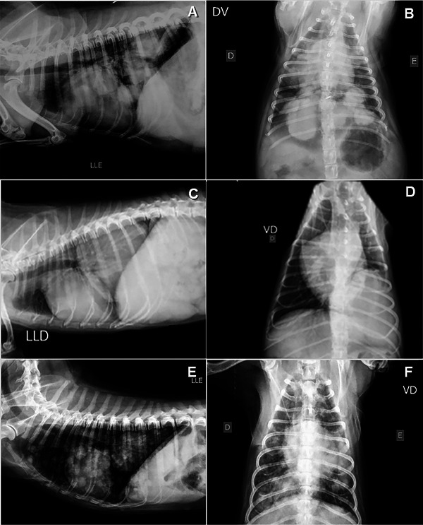
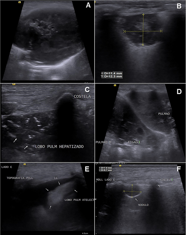
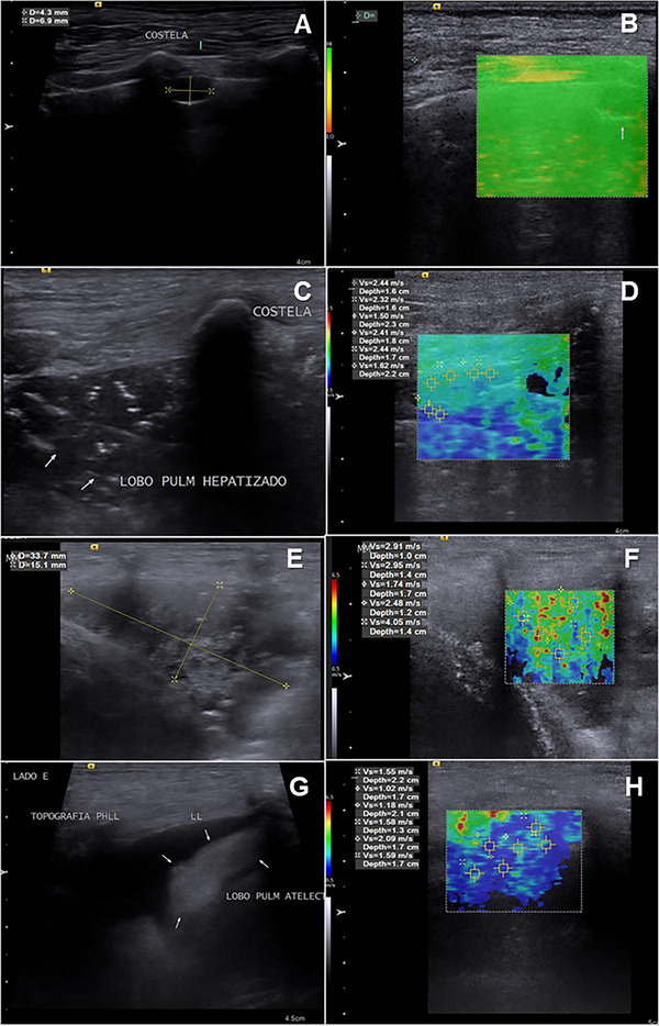
This study aimed to evaluate the use of acoustic radiation force impulse (ARFI) elastography as a diagnostic tool for lung lesions in dogs. Dogs referred to the Radiology Department of the Veterinary Teaching Hospital between 2020 and 2022 for the detection of lung lesions were included in the study. The characteristics of the lung lesions were assessed using radiography as a screening tool for localization, B-mode ultrasound for tissue characterization, and subsequently, both qualitative (elastogram grades 1-3) and quantitative (shear wave velocity-SWV) elastographic evaluations. The lesions were classified based on clinical, ultrasound, radiographic, histopathological, and/or cytological findings into the following categories: consolidations, atelectasis, or neoplasms (nodules and masses). Twenty-six dogs met the eligibility criteria and were included in the study. In some cases, the same dog had more than one type of lesion, resulting in the evaluation of 35 lung lesions: 13 masses, 8 nodules, 8 consolidations, and 4 areas of atelectasis. The quantitative elastographic evaluation revealed lower stiffness in atelectatic lesions (1.48 ± 0.35 m/s) compared with consolidations (2.94 ± 0.64 m/s), nodules (2.85 ± 1.40 m/s), and masses (3.13 ± 1.45 m/s), although no definitive diagnostic cut-off value was established, due to the limited number of benign lesions. The results suggest that ARFI elastography can be a valuable complementary tool alongside clinical data and conventional imaging techniques in assessing lung lesions in dogs. Future studies with a larger sample size of benign parenchymal lung lesions are needed to further explore the potential of elastography for predicting malignancy.
期刊介绍:
Veterinary Radiology & Ultrasound is a bimonthly, international, peer-reviewed, research journal devoted to the fields of veterinary diagnostic imaging and radiation oncology. Established in 1958, it is owned by the American College of Veterinary Radiology and is also the official journal for six affiliate veterinary organizations. Veterinary Radiology & Ultrasound is represented on the International Committee of Medical Journal Editors, World Association of Medical Editors, and Committee on Publication Ethics.
The mission of Veterinary Radiology & Ultrasound is to serve as a leading resource for high quality articles that advance scientific knowledge and standards of clinical practice in the areas of veterinary diagnostic radiology, computed tomography, magnetic resonance imaging, ultrasonography, nuclear imaging, radiation oncology, and interventional radiology. Manuscript types include original investigations, imaging diagnosis reports, review articles, editorials and letters to the Editor. Acceptance criteria include originality, significance, quality, reader interest, composition and adherence to author guidelines.

 求助内容:
求助内容: 应助结果提醒方式:


