Albert J. Rogers, Olga Reynbakh, Adnan Ahmed, Mina K. Chung, Rishi Charate, Hirad Yarmohammadi, Rakesh Gopinathannair, Hassan Khan, Dhanunjaya Lakkireddy, Miguel Leal, Uma Srivatsa, Natalia Trayanova, Elaine Y. Wan
{"title":"电生理学家的心血管成像技术。","authors":"Albert J. Rogers, Olga Reynbakh, Adnan Ahmed, Mina K. Chung, Rishi Charate, Hirad Yarmohammadi, Rakesh Gopinathannair, Hassan Khan, Dhanunjaya Lakkireddy, Miguel Leal, Uma Srivatsa, Natalia Trayanova, Elaine Y. Wan","doi":"10.1038/s44161-025-00648-8","DOIUrl":null,"url":null,"abstract":"Rapid technological advancements in noninvasive and invasive imaging including echocardiography, computed tomography, magnetic resonance imaging and positron emission tomography have allowed for improved anatomical visualization and precise measurement of cardiac structure and function. These imaging modalities allow for evaluation of how cardiac substrate changes, such as myocardial wall thickness, fibrosis, scarring and chamber enlargement and/or dilation, have an important role in arrhythmia initiation and perpetuation. Here, we review the various imaging techniques and modalities used by clinical and basic electrophysiologists to study cardiac arrhythmia mechanisms, periprocedural planning, risk stratification and precise delivery of ablation therapy. We also review the use of artificial intelligence and machine learning to improve identification of areas for triggered activity and isthmuses in reentrant arrhythmias, which may be favorable ablation targets. Rogers et al. review the current imaging modalities to investigate the mechanisms of cardiac arrhythmias and discuss the future impact of machine learning and artificial intelligence on digital imaging.","PeriodicalId":74245,"journal":{"name":"Nature cardiovascular research","volume":"4 5","pages":"514-525"},"PeriodicalIF":10.8000,"publicationDate":"2025-05-13","publicationTypes":"Journal Article","fieldsOfStudy":null,"isOpenAccess":false,"openAccessPdf":"","citationCount":"0","resultStr":"{\"title\":\"Cardiovascular imaging techniques for electrophysiologists\",\"authors\":\"Albert J. Rogers, Olga Reynbakh, Adnan Ahmed, Mina K. Chung, Rishi Charate, Hirad Yarmohammadi, Rakesh Gopinathannair, Hassan Khan, Dhanunjaya Lakkireddy, Miguel Leal, Uma Srivatsa, Natalia Trayanova, Elaine Y. Wan\",\"doi\":\"10.1038/s44161-025-00648-8\",\"DOIUrl\":null,\"url\":null,\"abstract\":\"Rapid technological advancements in noninvasive and invasive imaging including echocardiography, computed tomography, magnetic resonance imaging and positron emission tomography have allowed for improved anatomical visualization and precise measurement of cardiac structure and function. These imaging modalities allow for evaluation of how cardiac substrate changes, such as myocardial wall thickness, fibrosis, scarring and chamber enlargement and/or dilation, have an important role in arrhythmia initiation and perpetuation. Here, we review the various imaging techniques and modalities used by clinical and basic electrophysiologists to study cardiac arrhythmia mechanisms, periprocedural planning, risk stratification and precise delivery of ablation therapy. We also review the use of artificial intelligence and machine learning to improve identification of areas for triggered activity and isthmuses in reentrant arrhythmias, which may be favorable ablation targets. Rogers et al. review the current imaging modalities to investigate the mechanisms of cardiac arrhythmias and discuss the future impact of machine learning and artificial intelligence on digital imaging.\",\"PeriodicalId\":74245,\"journal\":{\"name\":\"Nature cardiovascular research\",\"volume\":\"4 5\",\"pages\":\"514-525\"},\"PeriodicalIF\":10.8000,\"publicationDate\":\"2025-05-13\",\"publicationTypes\":\"Journal Article\",\"fieldsOfStudy\":null,\"isOpenAccess\":false,\"openAccessPdf\":\"\",\"citationCount\":\"0\",\"resultStr\":null,\"platform\":\"Semanticscholar\",\"paperid\":null,\"PeriodicalName\":\"Nature cardiovascular research\",\"FirstCategoryId\":\"1085\",\"ListUrlMain\":\"https://www.nature.com/articles/s44161-025-00648-8\",\"RegionNum\":0,\"RegionCategory\":null,\"ArticlePicture\":[],\"TitleCN\":null,\"AbstractTextCN\":null,\"PMCID\":null,\"EPubDate\":\"\",\"PubModel\":\"\",\"JCR\":\"Q1\",\"JCRName\":\"CARDIAC & CARDIOVASCULAR SYSTEMS\",\"Score\":null,\"Total\":0}","platform":"Semanticscholar","paperid":null,"PeriodicalName":"Nature cardiovascular research","FirstCategoryId":"1085","ListUrlMain":"https://www.nature.com/articles/s44161-025-00648-8","RegionNum":0,"RegionCategory":null,"ArticlePicture":[],"TitleCN":null,"AbstractTextCN":null,"PMCID":null,"EPubDate":"","PubModel":"","JCR":"Q1","JCRName":"CARDIAC & CARDIOVASCULAR SYSTEMS","Score":null,"Total":0}
Cardiovascular imaging techniques for electrophysiologists
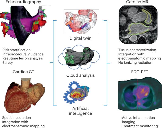
Rapid technological advancements in noninvasive and invasive imaging including echocardiography, computed tomography, magnetic resonance imaging and positron emission tomography have allowed for improved anatomical visualization and precise measurement of cardiac structure and function. These imaging modalities allow for evaluation of how cardiac substrate changes, such as myocardial wall thickness, fibrosis, scarring and chamber enlargement and/or dilation, have an important role in arrhythmia initiation and perpetuation. Here, we review the various imaging techniques and modalities used by clinical and basic electrophysiologists to study cardiac arrhythmia mechanisms, periprocedural planning, risk stratification and precise delivery of ablation therapy. We also review the use of artificial intelligence and machine learning to improve identification of areas for triggered activity and isthmuses in reentrant arrhythmias, which may be favorable ablation targets. Rogers et al. review the current imaging modalities to investigate the mechanisms of cardiac arrhythmias and discuss the future impact of machine learning and artificial intelligence on digital imaging.

 求助内容:
求助内容: 应助结果提醒方式:


