{"title":"阴道毛滴虫、白色念珠菌和crispr乳杆菌体外共培养模型:评估阴道炎中抗菌活性和微生物相互作用的系统。","authors":"Fernanda Gomes Cardoso, Luisa Trindade Dos Santos, Saulo Almeida Menezes, Graziela Vargas Rigo, Tiana Tasca","doi":"10.3389/fpara.2025.1523113","DOIUrl":null,"url":null,"abstract":"<p><p><i>Trichomonas vaginalis</i> is a flagellated protozoan causing trichomoniasis, the most common non-viral sexually transmitted infection. It is associated with various complications, particularly in asymptomatic carriers. Another major cause of vaginitis is <i>Candida albicans</i>, a normal member of the vaginal microbiota, which causes vulvovaginal candidiasis when immune imbalances occur, leading to recurrent infections. Treatment-resistant strains of these pathogens pose a significant challenge. <i>Lactobacillus crispatus</i>, a dominant species in the vaginal microbiota, produces antimicrobial compounds that help protect the vaginal mucosa. This study establishes an <i>in vitro</i> co-culture of <i>T. vaginalis</i>, <i>C. albicans</i>, and <i>L. crispatus</i> to simulate the vaginal microenvironment at the site of infection. MRS medium was chosen for the co-culture, with initial cell densities determined as follows: <i>T. vaginalis</i> at 1.0 × 10<sup>6</sup> trophozoites/mL (counted using a hemocytometer), 3.33 × 10<sup>4</sup> CFU/mL for <i>C. albicans</i>, and either 5.53 × 10<sup>6</sup> CFU/mL (for co-culture with the ATCC isolate) or 5.53 × 10<sup>7</sup> CFU/mL (for co-culture with a fresh clinical isolate) for <i>L. crispatus</i>. The cell densities of <i>C. albicans</i> and <i>L. crispatus</i> were quantified as colony-forming units (CFU) on selective agar. The incubation period for co-culture, ensuring optimal growth of all microorganisms, was 24 hours. In co-culture, <i>L. crispatus</i> at both tested densities acidified the medium. The co-culture system demonstrated lower MIC values for metronidazole (50 µM in the ATCC isolate co-culture and 25 µM with the fresh clinical isolate) and lower MFC values for fluconazole (6.25 µM), compared to monocultures of <i>T. vaginalis</i> (100 µM) and <i>C. albicans</i> (12.50 µM). Furthermore, the triple co-culture increased the cytotoxicity to vaginal cell and erythrocytes for the ATCC isolate while significantly inhibited both biofilm formation and metabolic activity of <i>C. albicans</i> (by up to 92% and 90%, respectively), as well as its yeast-to-hyphae transition (by up to 70%). SEM analyses highlighted the morphological differences among <i>T. vaginalis</i>, <i>C. albicans</i>, and <i>L. crispatus</i>, including isolate-specific size variations in the protozoan. These findings suggest that this <i>in vitro</i> co-culture system is a valuable tool for evaluating the antimicrobial efficacy of novel compounds against vaginitis pathogens and for studying interactions within the vaginal microenvironment.</p>","PeriodicalId":73098,"journal":{"name":"Frontiers in parasitology","volume":"4 ","pages":"1523113"},"PeriodicalIF":0.0000,"publicationDate":"2025-04-14","publicationTypes":"Journal Article","fieldsOfStudy":null,"isOpenAccess":false,"openAccessPdf":"https://www.ncbi.nlm.nih.gov/pmc/articles/PMC12034676/pdf/","citationCount":"0","resultStr":"{\"title\":\"<i>In vitro</i> co-culture model of <i>Trichomonas vaginalis</i>, <i>Candida albicans</i>, and <i>Lactobacillus crispatus</i>: a system for assessing antimicrobial activity and microorganism interactions in vaginitis.\",\"authors\":\"Fernanda Gomes Cardoso, Luisa Trindade Dos Santos, Saulo Almeida Menezes, Graziela Vargas Rigo, Tiana Tasca\",\"doi\":\"10.3389/fpara.2025.1523113\",\"DOIUrl\":null,\"url\":null,\"abstract\":\"<p><p><i>Trichomonas vaginalis</i> is a flagellated protozoan causing trichomoniasis, the most common non-viral sexually transmitted infection. It is associated with various complications, particularly in asymptomatic carriers. Another major cause of vaginitis is <i>Candida albicans</i>, a normal member of the vaginal microbiota, which causes vulvovaginal candidiasis when immune imbalances occur, leading to recurrent infections. Treatment-resistant strains of these pathogens pose a significant challenge. <i>Lactobacillus crispatus</i>, a dominant species in the vaginal microbiota, produces antimicrobial compounds that help protect the vaginal mucosa. This study establishes an <i>in vitro</i> co-culture of <i>T. vaginalis</i>, <i>C. albicans</i>, and <i>L. crispatus</i> to simulate the vaginal microenvironment at the site of infection. MRS medium was chosen for the co-culture, with initial cell densities determined as follows: <i>T. vaginalis</i> at 1.0 × 10<sup>6</sup> trophozoites/mL (counted using a hemocytometer), 3.33 × 10<sup>4</sup> CFU/mL for <i>C. albicans</i>, and either 5.53 × 10<sup>6</sup> CFU/mL (for co-culture with the ATCC isolate) or 5.53 × 10<sup>7</sup> CFU/mL (for co-culture with a fresh clinical isolate) for <i>L. crispatus</i>. The cell densities of <i>C. albicans</i> and <i>L. crispatus</i> were quantified as colony-forming units (CFU) on selective agar. The incubation period for co-culture, ensuring optimal growth of all microorganisms, was 24 hours. In co-culture, <i>L. crispatus</i> at both tested densities acidified the medium. The co-culture system demonstrated lower MIC values for metronidazole (50 µM in the ATCC isolate co-culture and 25 µM with the fresh clinical isolate) and lower MFC values for fluconazole (6.25 µM), compared to monocultures of <i>T. vaginalis</i> (100 µM) and <i>C. albicans</i> (12.50 µM). Furthermore, the triple co-culture increased the cytotoxicity to vaginal cell and erythrocytes for the ATCC isolate while significantly inhibited both biofilm formation and metabolic activity of <i>C. albicans</i> (by up to 92% and 90%, respectively), as well as its yeast-to-hyphae transition (by up to 70%). SEM analyses highlighted the morphological differences among <i>T. vaginalis</i>, <i>C. albicans</i>, and <i>L. crispatus</i>, including isolate-specific size variations in the protozoan. These findings suggest that this <i>in vitro</i> co-culture system is a valuable tool for evaluating the antimicrobial efficacy of novel compounds against vaginitis pathogens and for studying interactions within the vaginal microenvironment.</p>\",\"PeriodicalId\":73098,\"journal\":{\"name\":\"Frontiers in parasitology\",\"volume\":\"4 \",\"pages\":\"1523113\"},\"PeriodicalIF\":0.0000,\"publicationDate\":\"2025-04-14\",\"publicationTypes\":\"Journal Article\",\"fieldsOfStudy\":null,\"isOpenAccess\":false,\"openAccessPdf\":\"https://www.ncbi.nlm.nih.gov/pmc/articles/PMC12034676/pdf/\",\"citationCount\":\"0\",\"resultStr\":null,\"platform\":\"Semanticscholar\",\"paperid\":null,\"PeriodicalName\":\"Frontiers in parasitology\",\"FirstCategoryId\":\"1085\",\"ListUrlMain\":\"https://doi.org/10.3389/fpara.2025.1523113\",\"RegionNum\":0,\"RegionCategory\":null,\"ArticlePicture\":[],\"TitleCN\":null,\"AbstractTextCN\":null,\"PMCID\":null,\"EPubDate\":\"2025/1/1 0:00:00\",\"PubModel\":\"eCollection\",\"JCR\":\"\",\"JCRName\":\"\",\"Score\":null,\"Total\":0}","platform":"Semanticscholar","paperid":null,"PeriodicalName":"Frontiers in parasitology","FirstCategoryId":"1085","ListUrlMain":"https://doi.org/10.3389/fpara.2025.1523113","RegionNum":0,"RegionCategory":null,"ArticlePicture":[],"TitleCN":null,"AbstractTextCN":null,"PMCID":null,"EPubDate":"2025/1/1 0:00:00","PubModel":"eCollection","JCR":"","JCRName":"","Score":null,"Total":0}
In vitro co-culture model of Trichomonas vaginalis, Candida albicans, and Lactobacillus crispatus: a system for assessing antimicrobial activity and microorganism interactions in vaginitis.
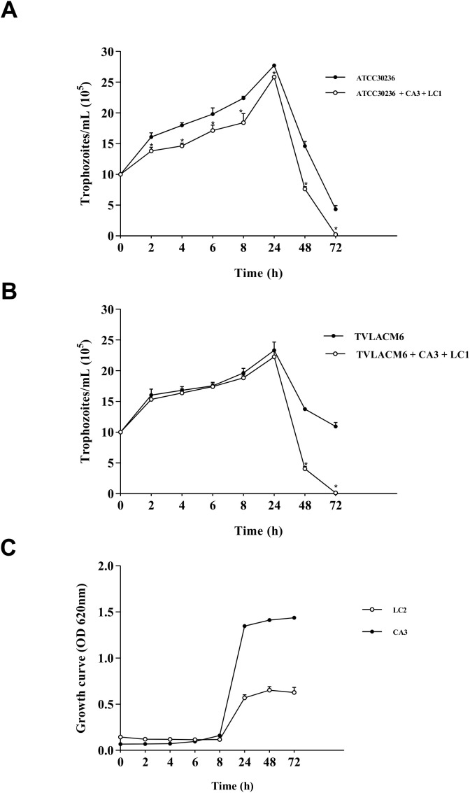
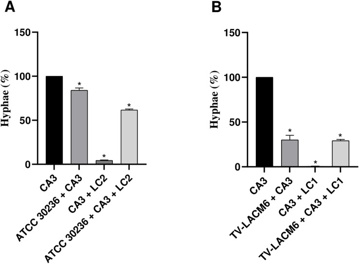
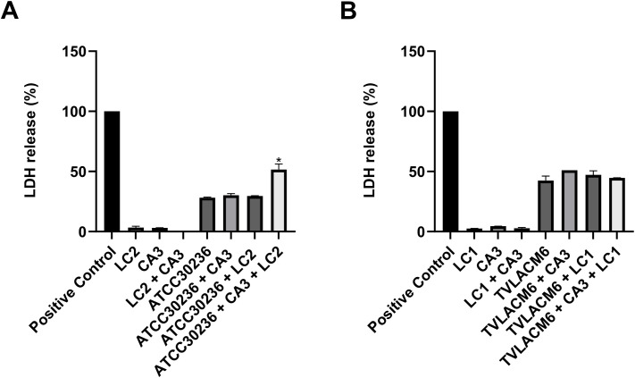
Trichomonas vaginalis is a flagellated protozoan causing trichomoniasis, the most common non-viral sexually transmitted infection. It is associated with various complications, particularly in asymptomatic carriers. Another major cause of vaginitis is Candida albicans, a normal member of the vaginal microbiota, which causes vulvovaginal candidiasis when immune imbalances occur, leading to recurrent infections. Treatment-resistant strains of these pathogens pose a significant challenge. Lactobacillus crispatus, a dominant species in the vaginal microbiota, produces antimicrobial compounds that help protect the vaginal mucosa. This study establishes an in vitro co-culture of T. vaginalis, C. albicans, and L. crispatus to simulate the vaginal microenvironment at the site of infection. MRS medium was chosen for the co-culture, with initial cell densities determined as follows: T. vaginalis at 1.0 × 10⁶ trophozoites/mL (counted using a hemocytometer), 3.33 × 10⁴ CFU/mL for C. albicans, and either 5.53 × 10⁶ CFU/mL (for co-culture with the ATCC isolate) or 5.53 × 10⁷ CFU/mL (for co-culture with a fresh clinical isolate) for L. crispatus. The cell densities of C. albicans and L. crispatus were quantified as colony-forming units (CFU) on selective agar. The incubation period for co-culture, ensuring optimal growth of all microorganisms, was 24 hours. In co-culture, L. crispatus at both tested densities acidified the medium. The co-culture system demonstrated lower MIC values for metronidazole (50 µM in the ATCC isolate co-culture and 25 µM with the fresh clinical isolate) and lower MFC values for fluconazole (6.25 µM), compared to monocultures of T. vaginalis (100 µM) and C. albicans (12.50 µM). Furthermore, the triple co-culture increased the cytotoxicity to vaginal cell and erythrocytes for the ATCC isolate while significantly inhibited both biofilm formation and metabolic activity of C. albicans (by up to 92% and 90%, respectively), as well as its yeast-to-hyphae transition (by up to 70%). SEM analyses highlighted the morphological differences among T. vaginalis, C. albicans, and L. crispatus, including isolate-specific size variations in the protozoan. These findings suggest that this in vitro co-culture system is a valuable tool for evaluating the antimicrobial efficacy of novel compounds against vaginitis pathogens and for studying interactions within the vaginal microenvironment.

 求助内容:
求助内容: 应助结果提醒方式:


