{"title":"磁共振血管造影诊断C5节段性椎动脉。","authors":"Kenji Takata, Midori Ueda, Kiyotaka Takeuchi, Hideaki Komiya, Daisuke Yoshikawa, Satomi Kanai, Tasuku Wakabayashi, Ayaki Kitano, Mariko Toyooka, Toyohiko Sakai, Tetsuya Tsujikawa","doi":"10.1007/s00276-025-03640-w","DOIUrl":null,"url":null,"abstract":"<p><strong>Purpose: </strong>The vertebral artery (VA) typically enters the subarachnoid space at the atlanto-occipital region. However, segmental variations can occur, with the VA entering the spinal canal at atypical levels. While a C3 segmental VA has been reported, no prior studies describe a C5 segmental VA. This case represents the first documented occurrence of this anomaly.</p><p><strong>Methods: </strong>An 8-year-old girl underwent brain magnetic resonance imaging for headache screening, which incidentally revealed an abnormal VA course.</p><p><strong>Results: </strong>Imaging revealed the absence of the left VA at the proximal V2 segment. Instead, a radiculomedullary artery at C4/5 entered the spinal canal and contributed to the formation of the anterior spinal artery (ASA), which ascended along the spinal cord. The right VA appeared normal; however, a radiculomedullary artery at the C3/4 level was identified, joining the contralateral radiculomedullary artery at the C1 level to form the ASA. Additionally, bilateral accessory middle cerebral arteries were observed. No clear association was found between this anomaly and the patient's headache, and she remained under observation.</p><p><strong>Conclusion: </strong>This anomaly may result from persistence of the fifth intersegmental artery. The vascular course resembled collateral circulation observed in acquired VA occlusion. Given its proximity to the spinal cord, potential risks include ischemic complications and spinal cord compression. This case highlights the importance of accurate imaging and careful surgical planning. Further studies on these rare vascular anomalies will enhance our understanding of VA variations and their clinical significance.</p>","PeriodicalId":49461,"journal":{"name":"Surgical and Radiologic Anatomy","volume":"47 1","pages":"132"},"PeriodicalIF":1.2000,"publicationDate":"2025-04-30","publicationTypes":"Journal Article","fieldsOfStudy":null,"isOpenAccess":false,"openAccessPdf":"https://www.ncbi.nlm.nih.gov/pmc/articles/PMC12043754/pdf/","citationCount":"0","resultStr":"{\"title\":\"Diagnosis of C5 segmental vertebral artery using magnetic resonance angiography.\",\"authors\":\"Kenji Takata, Midori Ueda, Kiyotaka Takeuchi, Hideaki Komiya, Daisuke Yoshikawa, Satomi Kanai, Tasuku Wakabayashi, Ayaki Kitano, Mariko Toyooka, Toyohiko Sakai, Tetsuya Tsujikawa\",\"doi\":\"10.1007/s00276-025-03640-w\",\"DOIUrl\":null,\"url\":null,\"abstract\":\"<p><strong>Purpose: </strong>The vertebral artery (VA) typically enters the subarachnoid space at the atlanto-occipital region. However, segmental variations can occur, with the VA entering the spinal canal at atypical levels. While a C3 segmental VA has been reported, no prior studies describe a C5 segmental VA. This case represents the first documented occurrence of this anomaly.</p><p><strong>Methods: </strong>An 8-year-old girl underwent brain magnetic resonance imaging for headache screening, which incidentally revealed an abnormal VA course.</p><p><strong>Results: </strong>Imaging revealed the absence of the left VA at the proximal V2 segment. Instead, a radiculomedullary artery at C4/5 entered the spinal canal and contributed to the formation of the anterior spinal artery (ASA), which ascended along the spinal cord. The right VA appeared normal; however, a radiculomedullary artery at the C3/4 level was identified, joining the contralateral radiculomedullary artery at the C1 level to form the ASA. Additionally, bilateral accessory middle cerebral arteries were observed. No clear association was found between this anomaly and the patient's headache, and she remained under observation.</p><p><strong>Conclusion: </strong>This anomaly may result from persistence of the fifth intersegmental artery. The vascular course resembled collateral circulation observed in acquired VA occlusion. Given its proximity to the spinal cord, potential risks include ischemic complications and spinal cord compression. This case highlights the importance of accurate imaging and careful surgical planning. Further studies on these rare vascular anomalies will enhance our understanding of VA variations and their clinical significance.</p>\",\"PeriodicalId\":49461,\"journal\":{\"name\":\"Surgical and Radiologic Anatomy\",\"volume\":\"47 1\",\"pages\":\"132\"},\"PeriodicalIF\":1.2000,\"publicationDate\":\"2025-04-30\",\"publicationTypes\":\"Journal Article\",\"fieldsOfStudy\":null,\"isOpenAccess\":false,\"openAccessPdf\":\"https://www.ncbi.nlm.nih.gov/pmc/articles/PMC12043754/pdf/\",\"citationCount\":\"0\",\"resultStr\":null,\"platform\":\"Semanticscholar\",\"paperid\":null,\"PeriodicalName\":\"Surgical and Radiologic Anatomy\",\"FirstCategoryId\":\"3\",\"ListUrlMain\":\"https://doi.org/10.1007/s00276-025-03640-w\",\"RegionNum\":4,\"RegionCategory\":\"医学\",\"ArticlePicture\":[],\"TitleCN\":null,\"AbstractTextCN\":null,\"PMCID\":null,\"EPubDate\":\"\",\"PubModel\":\"\",\"JCR\":\"Q2\",\"JCRName\":\"Medicine\",\"Score\":null,\"Total\":0}","platform":"Semanticscholar","paperid":null,"PeriodicalName":"Surgical and Radiologic Anatomy","FirstCategoryId":"3","ListUrlMain":"https://doi.org/10.1007/s00276-025-03640-w","RegionNum":4,"RegionCategory":"医学","ArticlePicture":[],"TitleCN":null,"AbstractTextCN":null,"PMCID":null,"EPubDate":"","PubModel":"","JCR":"Q2","JCRName":"Medicine","Score":null,"Total":0}
Diagnosis of C5 segmental vertebral artery using magnetic resonance angiography.
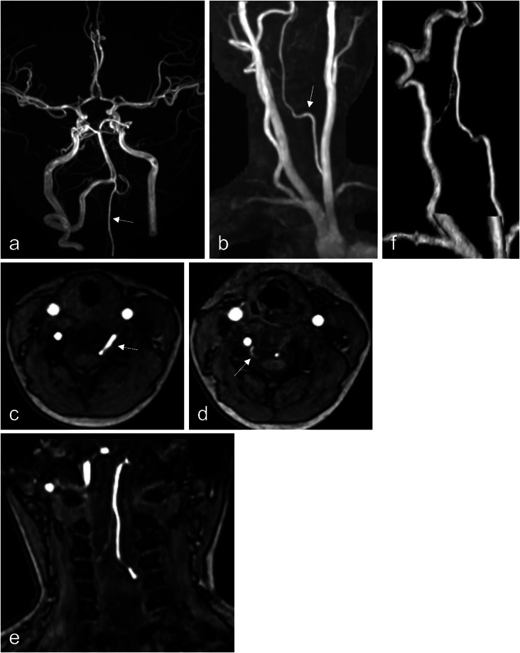
Purpose: The vertebral artery (VA) typically enters the subarachnoid space at the atlanto-occipital region. However, segmental variations can occur, with the VA entering the spinal canal at atypical levels. While a C3 segmental VA has been reported, no prior studies describe a C5 segmental VA. This case represents the first documented occurrence of this anomaly.
Methods: An 8-year-old girl underwent brain magnetic resonance imaging for headache screening, which incidentally revealed an abnormal VA course.
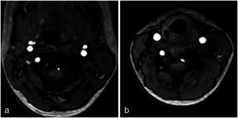
Results: Imaging revealed the absence of the left VA at the proximal V2 segment. Instead, a radiculomedullary artery at C4/5 entered the spinal canal and contributed to the formation of the anterior spinal artery (ASA), which ascended along the spinal cord. The right VA appeared normal; however, a radiculomedullary artery at the C3/4 level was identified, joining the contralateral radiculomedullary artery at the C1 level to form the ASA. Additionally, bilateral accessory middle cerebral arteries were observed. No clear association was found between this anomaly and the patient's headache, and she remained under observation.
Conclusion: This anomaly may result from persistence of the fifth intersegmental artery. The vascular course resembled collateral circulation observed in acquired VA occlusion. Given its proximity to the spinal cord, potential risks include ischemic complications and spinal cord compression. This case highlights the importance of accurate imaging and careful surgical planning. Further studies on these rare vascular anomalies will enhance our understanding of VA variations and their clinical significance.
期刊介绍:
Anatomy is a morphological science which cannot fail to interest the clinician. The practical application of anatomical research to clinical problems necessitates special adaptation and selectivity in choosing from numerous international works. Although there is a tendency to believe that meaningful advances in anatomy are unlikely, constant revision is necessary. Surgical and Radiologic Anatomy, the first international journal of Clinical anatomy has been created in this spirit.
Its goal is to serve clinicians, regardless of speciality-physicians, surgeons, radiologists or other specialists-as an indispensable aid with which they can improve their knowledge of anatomy. Each issue includes: Original papers, review articles, articles on the anatomical bases of medical, surgical and radiological techniques, articles of normal radiologic anatomy, brief reviews of anatomical publications of clinical interest.
Particular attention is given to high quality illustrations, which are indispensable for a better understanding of anatomical problems.
Surgical and Radiologic Anatomy is a journal written by anatomists for clinicians with a special interest in anatomy.

 求助内容:
求助内容: 应助结果提醒方式:


