{"title":"脑岛解剖定位的数学和动态建模。","authors":"Eren Ogut","doi":"10.1007/s12021-025-09727-4","DOIUrl":null,"url":null,"abstract":"<p><p>The insula, a deeply situated cortical structure beneath the Sylvian sulcus, plays a critical role in sensory integration, emotion regulation, and cognitive control in the brain. Although several studies have described its anatomical and functional characteristics, mathematical models that quantitatively represent the insula's complex structure and connectivity are lacking. This study aimed to develop a mathematical model to represent the anatomical localization and functional organization of the insula, drawing on current neuroimaging findings and established anatomical data. A three-dimensional (3D) ellipsoid model was constructed to mathematically represent the anatomical boundaries of the insula using Montreal Neurological Institute (MNI) coordinate data. This geometric model adapts the ellipsoid equation to reflect the spatial configuration of the insula and is primarily based on cytoarchitectonic mapping and anatomical literature. Relevant findings from prior imaging research, particularly those reporting microstructural variations across insular subdivisions, were reviewed and conceptually integrated to guide the model's structural assumptions and interpretation of potential applications. The ellipsoid-based 3D model accurately represented the anatomical dimensions and spatial localization of the right insula, centered at the MNI coordinates (40, 5, 5 mm), and matched well with the known volumetric data. Functional regions (face, hand, and foot) were successfully plotted within the model, and statistical analysis confirmed significant differences along the anteroposterior and superoinferior axes (p < 0.01 and p < 0.05, respectively). Dynamic simulations revealed oscillatory patterns of excitatory and inhibitory neural activity, consistent with established insular neurophysiology. Additionally, connectivity modeling demonstrated strong bidirectional interactions between the insula and key regions, such as the prefrontal cortex and anterior cingulate cortex (ACC), reflecting its integrative role in brain networks. This study presents a scientifically validated mathematical model that captures the anatomical structure, functional subdivisions, and dynamic connectivity patterns of the insula. By integrating anatomical data with computational simulations, this model provides a foundation for future research in neuroimaging, functional mapping, and clinical applications involving insula-related disorders.</p>","PeriodicalId":49761,"journal":{"name":"Neuroinformatics","volume":"23 2","pages":"29"},"PeriodicalIF":3.1000,"publicationDate":"2025-04-23","publicationTypes":"Journal Article","fieldsOfStudy":null,"isOpenAccess":false,"openAccessPdf":"https://www.ncbi.nlm.nih.gov/pmc/articles/PMC12018515/pdf/","citationCount":"0","resultStr":"{\"title\":\"Mathematical and Dynamic Modeling of the Anatomical Localization of the Insula in the Brain.\",\"authors\":\"Eren Ogut\",\"doi\":\"10.1007/s12021-025-09727-4\",\"DOIUrl\":null,\"url\":null,\"abstract\":\"<p><p>The insula, a deeply situated cortical structure beneath the Sylvian sulcus, plays a critical role in sensory integration, emotion regulation, and cognitive control in the brain. Although several studies have described its anatomical and functional characteristics, mathematical models that quantitatively represent the insula's complex structure and connectivity are lacking. This study aimed to develop a mathematical model to represent the anatomical localization and functional organization of the insula, drawing on current neuroimaging findings and established anatomical data. A three-dimensional (3D) ellipsoid model was constructed to mathematically represent the anatomical boundaries of the insula using Montreal Neurological Institute (MNI) coordinate data. This geometric model adapts the ellipsoid equation to reflect the spatial configuration of the insula and is primarily based on cytoarchitectonic mapping and anatomical literature. Relevant findings from prior imaging research, particularly those reporting microstructural variations across insular subdivisions, were reviewed and conceptually integrated to guide the model's structural assumptions and interpretation of potential applications. The ellipsoid-based 3D model accurately represented the anatomical dimensions and spatial localization of the right insula, centered at the MNI coordinates (40, 5, 5 mm), and matched well with the known volumetric data. Functional regions (face, hand, and foot) were successfully plotted within the model, and statistical analysis confirmed significant differences along the anteroposterior and superoinferior axes (p < 0.01 and p < 0.05, respectively). Dynamic simulations revealed oscillatory patterns of excitatory and inhibitory neural activity, consistent with established insular neurophysiology. Additionally, connectivity modeling demonstrated strong bidirectional interactions between the insula and key regions, such as the prefrontal cortex and anterior cingulate cortex (ACC), reflecting its integrative role in brain networks. This study presents a scientifically validated mathematical model that captures the anatomical structure, functional subdivisions, and dynamic connectivity patterns of the insula. By integrating anatomical data with computational simulations, this model provides a foundation for future research in neuroimaging, functional mapping, and clinical applications involving insula-related disorders.</p>\",\"PeriodicalId\":49761,\"journal\":{\"name\":\"Neuroinformatics\",\"volume\":\"23 2\",\"pages\":\"29\"},\"PeriodicalIF\":3.1000,\"publicationDate\":\"2025-04-23\",\"publicationTypes\":\"Journal Article\",\"fieldsOfStudy\":null,\"isOpenAccess\":false,\"openAccessPdf\":\"https://www.ncbi.nlm.nih.gov/pmc/articles/PMC12018515/pdf/\",\"citationCount\":\"0\",\"resultStr\":null,\"platform\":\"Semanticscholar\",\"paperid\":null,\"PeriodicalName\":\"Neuroinformatics\",\"FirstCategoryId\":\"3\",\"ListUrlMain\":\"https://doi.org/10.1007/s12021-025-09727-4\",\"RegionNum\":4,\"RegionCategory\":\"医学\",\"ArticlePicture\":[],\"TitleCN\":null,\"AbstractTextCN\":null,\"PMCID\":null,\"EPubDate\":\"\",\"PubModel\":\"\",\"JCR\":\"Q2\",\"JCRName\":\"COMPUTER SCIENCE, INTERDISCIPLINARY APPLICATIONS\",\"Score\":null,\"Total\":0}","platform":"Semanticscholar","paperid":null,"PeriodicalName":"Neuroinformatics","FirstCategoryId":"3","ListUrlMain":"https://doi.org/10.1007/s12021-025-09727-4","RegionNum":4,"RegionCategory":"医学","ArticlePicture":[],"TitleCN":null,"AbstractTextCN":null,"PMCID":null,"EPubDate":"","PubModel":"","JCR":"Q2","JCRName":"COMPUTER SCIENCE, INTERDISCIPLINARY APPLICATIONS","Score":null,"Total":0}
Mathematical and Dynamic Modeling of the Anatomical Localization of the Insula in the Brain.
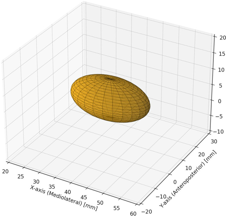
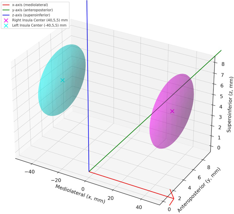
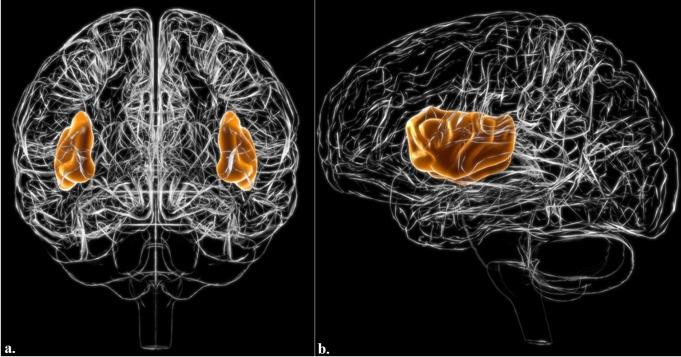
The insula, a deeply situated cortical structure beneath the Sylvian sulcus, plays a critical role in sensory integration, emotion regulation, and cognitive control in the brain. Although several studies have described its anatomical and functional characteristics, mathematical models that quantitatively represent the insula's complex structure and connectivity are lacking. This study aimed to develop a mathematical model to represent the anatomical localization and functional organization of the insula, drawing on current neuroimaging findings and established anatomical data. A three-dimensional (3D) ellipsoid model was constructed to mathematically represent the anatomical boundaries of the insula using Montreal Neurological Institute (MNI) coordinate data. This geometric model adapts the ellipsoid equation to reflect the spatial configuration of the insula and is primarily based on cytoarchitectonic mapping and anatomical literature. Relevant findings from prior imaging research, particularly those reporting microstructural variations across insular subdivisions, were reviewed and conceptually integrated to guide the model's structural assumptions and interpretation of potential applications. The ellipsoid-based 3D model accurately represented the anatomical dimensions and spatial localization of the right insula, centered at the MNI coordinates (40, 5, 5 mm), and matched well with the known volumetric data. Functional regions (face, hand, and foot) were successfully plotted within the model, and statistical analysis confirmed significant differences along the anteroposterior and superoinferior axes (p < 0.01 and p < 0.05, respectively). Dynamic simulations revealed oscillatory patterns of excitatory and inhibitory neural activity, consistent with established insular neurophysiology. Additionally, connectivity modeling demonstrated strong bidirectional interactions between the insula and key regions, such as the prefrontal cortex and anterior cingulate cortex (ACC), reflecting its integrative role in brain networks. This study presents a scientifically validated mathematical model that captures the anatomical structure, functional subdivisions, and dynamic connectivity patterns of the insula. By integrating anatomical data with computational simulations, this model provides a foundation for future research in neuroimaging, functional mapping, and clinical applications involving insula-related disorders.
期刊介绍:
Neuroinformatics publishes original articles and reviews with an emphasis on data structure and software tools related to analysis, modeling, integration, and sharing in all areas of neuroscience research. The editors particularly invite contributions on: (1) Theory and methodology, including discussions on ontologies, modeling approaches, database design, and meta-analyses; (2) Descriptions of developed databases and software tools, and of the methods for their distribution; (3) Relevant experimental results, such as reports accompanie by the release of massive data sets; (4) Computational simulations of models integrating and organizing complex data; and (5) Neuroengineering approaches, including hardware, robotics, and information theory studies.
 求助内容:
求助内容: 应助结果提醒方式:


