Rita M Jalkh, Yara Yammine, Nader Zalaquett, Houssein Darwish, Zeina Korban
{"title":"双侧鼻内窥镜视神经减压术治疗婴儿骨质疏松1例。","authors":"Rita M Jalkh, Yara Yammine, Nader Zalaquett, Houssein Darwish, Zeina Korban","doi":"10.1055/a-2554-2426","DOIUrl":null,"url":null,"abstract":"<p><strong>Background: </strong>Osteopetrosis is a rare genetic disorder characterized by abnormal bone density and structure, often leading to vision loss due to optic canal stenosis and consequent nerve compression. Early intervention is critical to prevent irreversible damage. This case report discusses the management of bilateral optic nerve compression in an infant with osteopetrosis.</p><p><strong>Case description: </strong>A 7-month-old male with a family history of osteopetrosis presented with hepatosplenomegaly. The infant was diagnosed with osteopetrosis based on radiological findings and genetic testing. Ophthalmologic examination and magnetic resonance imaging showed evidence of bilateral optic nerve compression. Endoscopic transcaruncular optic nerve decompression was not attainable The patient underwent a bilateral expanded endoscopic endonasal medial orbital wall and optic canal decompression.</p><p><strong>Conclusion: </strong>This is one of the few reported cases of endoscopic endonasal optic nerve decompression surgery on an infant. Endoscopic endonasal optic nerve decompression surgery is a viable and effective treatment option for optic nerve compression in infants with osteopetrosis, especially in cases where cost of surgery is a limiting factor for patients. This approach provides direct access to the optic canal with minimal morbidity, offering significant potential for visual recovery, and an improved quality of life. Our patient represents the youngest reported infant in the literature, demonstrating the potential for undergoing this surgical approach at the earliest possible age to aid with his prognosis.</p>","PeriodicalId":44256,"journal":{"name":"Journal of Neurological Surgery Reports","volume":"86 2","pages":"e65-e71"},"PeriodicalIF":0.7000,"publicationDate":"2025-04-11","publicationTypes":"Journal Article","fieldsOfStudy":null,"isOpenAccess":false,"openAccessPdf":"https://www.ncbi.nlm.nih.gov/pmc/articles/PMC12020546/pdf/","citationCount":"0","resultStr":"{\"title\":\"Bilateral Endoscopic Endonasal Optic Nerve Decompression in an Infant with Osteopetrosis: A Case Report.\",\"authors\":\"Rita M Jalkh, Yara Yammine, Nader Zalaquett, Houssein Darwish, Zeina Korban\",\"doi\":\"10.1055/a-2554-2426\",\"DOIUrl\":null,\"url\":null,\"abstract\":\"<p><strong>Background: </strong>Osteopetrosis is a rare genetic disorder characterized by abnormal bone density and structure, often leading to vision loss due to optic canal stenosis and consequent nerve compression. Early intervention is critical to prevent irreversible damage. This case report discusses the management of bilateral optic nerve compression in an infant with osteopetrosis.</p><p><strong>Case description: </strong>A 7-month-old male with a family history of osteopetrosis presented with hepatosplenomegaly. The infant was diagnosed with osteopetrosis based on radiological findings and genetic testing. Ophthalmologic examination and magnetic resonance imaging showed evidence of bilateral optic nerve compression. Endoscopic transcaruncular optic nerve decompression was not attainable The patient underwent a bilateral expanded endoscopic endonasal medial orbital wall and optic canal decompression.</p><p><strong>Conclusion: </strong>This is one of the few reported cases of endoscopic endonasal optic nerve decompression surgery on an infant. Endoscopic endonasal optic nerve decompression surgery is a viable and effective treatment option for optic nerve compression in infants with osteopetrosis, especially in cases where cost of surgery is a limiting factor for patients. This approach provides direct access to the optic canal with minimal morbidity, offering significant potential for visual recovery, and an improved quality of life. Our patient represents the youngest reported infant in the literature, demonstrating the potential for undergoing this surgical approach at the earliest possible age to aid with his prognosis.</p>\",\"PeriodicalId\":44256,\"journal\":{\"name\":\"Journal of Neurological Surgery Reports\",\"volume\":\"86 2\",\"pages\":\"e65-e71\"},\"PeriodicalIF\":0.7000,\"publicationDate\":\"2025-04-11\",\"publicationTypes\":\"Journal Article\",\"fieldsOfStudy\":null,\"isOpenAccess\":false,\"openAccessPdf\":\"https://www.ncbi.nlm.nih.gov/pmc/articles/PMC12020546/pdf/\",\"citationCount\":\"0\",\"resultStr\":null,\"platform\":\"Semanticscholar\",\"paperid\":null,\"PeriodicalName\":\"Journal of Neurological Surgery Reports\",\"FirstCategoryId\":\"1085\",\"ListUrlMain\":\"https://doi.org/10.1055/a-2554-2426\",\"RegionNum\":0,\"RegionCategory\":null,\"ArticlePicture\":[],\"TitleCN\":null,\"AbstractTextCN\":null,\"PMCID\":null,\"EPubDate\":\"2025/4/1 0:00:00\",\"PubModel\":\"eCollection\",\"JCR\":\"Q4\",\"JCRName\":\"CLINICAL NEUROLOGY\",\"Score\":null,\"Total\":0}","platform":"Semanticscholar","paperid":null,"PeriodicalName":"Journal of Neurological Surgery Reports","FirstCategoryId":"1085","ListUrlMain":"https://doi.org/10.1055/a-2554-2426","RegionNum":0,"RegionCategory":null,"ArticlePicture":[],"TitleCN":null,"AbstractTextCN":null,"PMCID":null,"EPubDate":"2025/4/1 0:00:00","PubModel":"eCollection","JCR":"Q4","JCRName":"CLINICAL NEUROLOGY","Score":null,"Total":0}
Bilateral Endoscopic Endonasal Optic Nerve Decompression in an Infant with Osteopetrosis: A Case Report.
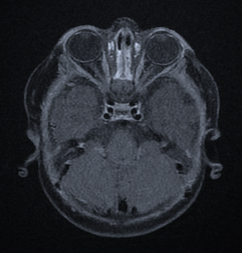
Background: Osteopetrosis is a rare genetic disorder characterized by abnormal bone density and structure, often leading to vision loss due to optic canal stenosis and consequent nerve compression. Early intervention is critical to prevent irreversible damage. This case report discusses the management of bilateral optic nerve compression in an infant with osteopetrosis.
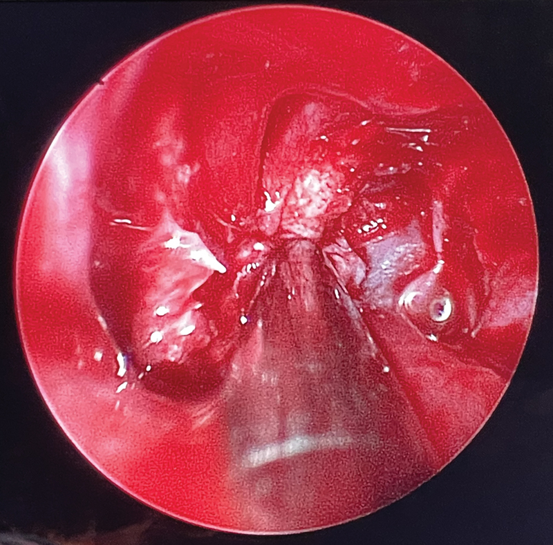
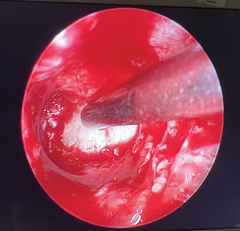
Case description: A 7-month-old male with a family history of osteopetrosis presented with hepatosplenomegaly. The infant was diagnosed with osteopetrosis based on radiological findings and genetic testing. Ophthalmologic examination and magnetic resonance imaging showed evidence of bilateral optic nerve compression. Endoscopic transcaruncular optic nerve decompression was not attainable The patient underwent a bilateral expanded endoscopic endonasal medial orbital wall and optic canal decompression.
Conclusion: This is one of the few reported cases of endoscopic endonasal optic nerve decompression surgery on an infant. Endoscopic endonasal optic nerve decompression surgery is a viable and effective treatment option for optic nerve compression in infants with osteopetrosis, especially in cases where cost of surgery is a limiting factor for patients. This approach provides direct access to the optic canal with minimal morbidity, offering significant potential for visual recovery, and an improved quality of life. Our patient represents the youngest reported infant in the literature, demonstrating the potential for undergoing this surgical approach at the earliest possible age to aid with his prognosis.

 求助内容:
求助内容: 应助结果提醒方式:


