{"title":"肠替代造影剂排泄对失蛋白性肠病犬的延迟计算机断层扫描。","authors":"Yujin Lee, Hojung Choi, Young-Won Lee, Kija Lee, Sooyoung Choi","doi":"10.1111/vru.70039","DOIUrl":null,"url":null,"abstract":"<p><p>The intestinal vicarious contrast medium excretion (VCME) can occur in dogs with protein-losing enteropathy (PLE), and studies for intestinal VCME in dogs are lacking. This retrospective case-control study aimed to assess whether intestinal VCME could be observed on delayed CT in dogs with and without PLE. Thirty dogs who underwent abdominal delayed CT in the 10 min-delayed phase following the injection of contrast medium were enrolled. Six dogs were classified into the group with enteropathy based on imaging findings or abnormal results from cytology or histology. The six dogs had concurrent hypoalbuminemia and were diagnosed with presumed PLE. Five of the six dogs in the group with enteropathy had intestinal VCME. In the 24 dogs of the group without enteropathy, intestinal VCME was not detected on delayed CT, and VCME to the cisterna chyli was observed in one dog. The frequency of intestinal VCME was significantly higher in the group with enteropathy than in the group without enteropathy (p < .001). The presence of intestinal VCME on the delayed CT can be observed in dogs with PLE, and it can be used as noninvasive additional supportive evidence of canine PLE prior to histopathologic evaluation.</p>","PeriodicalId":23581,"journal":{"name":"Veterinary Radiology & Ultrasound","volume":"66 3","pages":"e70039"},"PeriodicalIF":1.5000,"publicationDate":"2025-05-01","publicationTypes":"Journal Article","fieldsOfStudy":null,"isOpenAccess":false,"openAccessPdf":"https://www.ncbi.nlm.nih.gov/pmc/articles/PMC12059201/pdf/","citationCount":"0","resultStr":"{\"title\":\"Intestinal Vicarious Contrast Medium Excretion on Delayed Computed Tomography in Dogs with Protein-Losing Enteropathy.\",\"authors\":\"Yujin Lee, Hojung Choi, Young-Won Lee, Kija Lee, Sooyoung Choi\",\"doi\":\"10.1111/vru.70039\",\"DOIUrl\":null,\"url\":null,\"abstract\":\"<p><p>The intestinal vicarious contrast medium excretion (VCME) can occur in dogs with protein-losing enteropathy (PLE), and studies for intestinal VCME in dogs are lacking. This retrospective case-control study aimed to assess whether intestinal VCME could be observed on delayed CT in dogs with and without PLE. Thirty dogs who underwent abdominal delayed CT in the 10 min-delayed phase following the injection of contrast medium were enrolled. Six dogs were classified into the group with enteropathy based on imaging findings or abnormal results from cytology or histology. The six dogs had concurrent hypoalbuminemia and were diagnosed with presumed PLE. Five of the six dogs in the group with enteropathy had intestinal VCME. In the 24 dogs of the group without enteropathy, intestinal VCME was not detected on delayed CT, and VCME to the cisterna chyli was observed in one dog. The frequency of intestinal VCME was significantly higher in the group with enteropathy than in the group without enteropathy (p < .001). The presence of intestinal VCME on the delayed CT can be observed in dogs with PLE, and it can be used as noninvasive additional supportive evidence of canine PLE prior to histopathologic evaluation.</p>\",\"PeriodicalId\":23581,\"journal\":{\"name\":\"Veterinary Radiology & Ultrasound\",\"volume\":\"66 3\",\"pages\":\"e70039\"},\"PeriodicalIF\":1.5000,\"publicationDate\":\"2025-05-01\",\"publicationTypes\":\"Journal Article\",\"fieldsOfStudy\":null,\"isOpenAccess\":false,\"openAccessPdf\":\"https://www.ncbi.nlm.nih.gov/pmc/articles/PMC12059201/pdf/\",\"citationCount\":\"0\",\"resultStr\":null,\"platform\":\"Semanticscholar\",\"paperid\":null,\"PeriodicalName\":\"Veterinary Radiology & Ultrasound\",\"FirstCategoryId\":\"97\",\"ListUrlMain\":\"https://doi.org/10.1111/vru.70039\",\"RegionNum\":2,\"RegionCategory\":\"农林科学\",\"ArticlePicture\":[],\"TitleCN\":null,\"AbstractTextCN\":null,\"PMCID\":null,\"EPubDate\":\"\",\"PubModel\":\"\",\"JCR\":\"Q2\",\"JCRName\":\"VETERINARY SCIENCES\",\"Score\":null,\"Total\":0}","platform":"Semanticscholar","paperid":null,"PeriodicalName":"Veterinary Radiology & Ultrasound","FirstCategoryId":"97","ListUrlMain":"https://doi.org/10.1111/vru.70039","RegionNum":2,"RegionCategory":"农林科学","ArticlePicture":[],"TitleCN":null,"AbstractTextCN":null,"PMCID":null,"EPubDate":"","PubModel":"","JCR":"Q2","JCRName":"VETERINARY SCIENCES","Score":null,"Total":0}
Intestinal Vicarious Contrast Medium Excretion on Delayed Computed Tomography in Dogs with Protein-Losing Enteropathy.
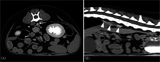
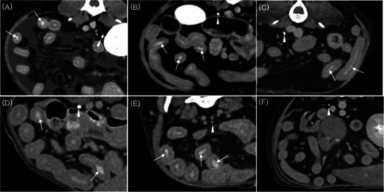
The intestinal vicarious contrast medium excretion (VCME) can occur in dogs with protein-losing enteropathy (PLE), and studies for intestinal VCME in dogs are lacking. This retrospective case-control study aimed to assess whether intestinal VCME could be observed on delayed CT in dogs with and without PLE. Thirty dogs who underwent abdominal delayed CT in the 10 min-delayed phase following the injection of contrast medium were enrolled. Six dogs were classified into the group with enteropathy based on imaging findings or abnormal results from cytology or histology. The six dogs had concurrent hypoalbuminemia and were diagnosed with presumed PLE. Five of the six dogs in the group with enteropathy had intestinal VCME. In the 24 dogs of the group without enteropathy, intestinal VCME was not detected on delayed CT, and VCME to the cisterna chyli was observed in one dog. The frequency of intestinal VCME was significantly higher in the group with enteropathy than in the group without enteropathy (p < .001). The presence of intestinal VCME on the delayed CT can be observed in dogs with PLE, and it can be used as noninvasive additional supportive evidence of canine PLE prior to histopathologic evaluation.
期刊介绍:
Veterinary Radiology & Ultrasound is a bimonthly, international, peer-reviewed, research journal devoted to the fields of veterinary diagnostic imaging and radiation oncology. Established in 1958, it is owned by the American College of Veterinary Radiology and is also the official journal for six affiliate veterinary organizations. Veterinary Radiology & Ultrasound is represented on the International Committee of Medical Journal Editors, World Association of Medical Editors, and Committee on Publication Ethics.
The mission of Veterinary Radiology & Ultrasound is to serve as a leading resource for high quality articles that advance scientific knowledge and standards of clinical practice in the areas of veterinary diagnostic radiology, computed tomography, magnetic resonance imaging, ultrasonography, nuclear imaging, radiation oncology, and interventional radiology. Manuscript types include original investigations, imaging diagnosis reports, review articles, editorials and letters to the Editor. Acceptance criteria include originality, significance, quality, reader interest, composition and adherence to author guidelines.

 求助内容:
求助内容: 应助结果提醒方式:


