Enayat Darabi, Seyed Mahmoud Sadjjadi, Tahereh Mohammadzadeh, Mehdi Karamian, Seyed Mohammad Owji, Bahareh Sedaghat
{"title":"伊朗设拉子地区人囊性棘球蚴病的严格感细粒棘球绦虫和加拿大棘球绦虫基因型的组织病理学差异","authors":"Enayat Darabi, Seyed Mahmoud Sadjjadi, Tahereh Mohammadzadeh, Mehdi Karamian, Seyed Mohammad Owji, Bahareh Sedaghat","doi":"10.18502/ijpa.v20i1.18102","DOIUrl":null,"url":null,"abstract":"<p><strong>Background: </strong>We aimed to determine the genotypes of <i>Echinococcus granulosus sensu lato</i> (<i>s.l.</i>) using DNA regions within the <i>NADH dehydrogenase subunit-1</i> (<i>nad1</i>) mitochondrial genes in formalin-fixed paraffin-embedded (FFPE) isolates of human cystic echinococcosis cysts (CE cysts) and compare their histopathologic alterations.</p><p><strong>Methods: </strong>Out of 135 samples, 21 high-quality PCR positive samples were selected for sequencing and were deposited into GenBank database. Moreover, histopathological changes of <i>E. granulosus sensu stricto</i> (G1 genotype) and <i>E. canadensis</i> (G6 genotype) cases were also compared.</p><p><strong>Results: </strong>Based on the sequencing results, 16 cases were diagnosed as <i>E. granulosus s.s.</i> (G1-G3 genotype) and 5 cases as <i>E. canadensis</i> (G6 genotype). Five haplotypes of <i>E. granulosus</i> were identified from 21 <i>nad1</i> sequences. The histopathological alterations in both genotypes showed laminated layer of CE without inflammatory cells. In a few cases of the G6 genotype, neutrophils in the outer cuticular layer with mild vascular and congestion were observed. Cell debris with multiple areas of necrosis, as well as scanty lymphoplasma cells in the outer cuticular layer were observed in G1-G3 genotype cases. So, the histopathological differences between the two genotypes are not noticeable enough to be differentiated by microscopical observations.</p><p><strong>Conclusion: </strong><i>E. granulosus s.s.</i> (G1-G3) and <i>E. canadensis</i> (G6 genotype) are prevalent among CE patients. In general, five haplotypes were identified by <i>nad1</i> genes analysis. The histopathological differences between the two genotypes have not been so big to be differentiated by microscopic observations.</p>","PeriodicalId":14669,"journal":{"name":"Iranian Journal of Parasitology","volume":"20 1","pages":"21-31"},"PeriodicalIF":0.9000,"publicationDate":"2025-01-01","publicationTypes":"Journal Article","fieldsOfStudy":null,"isOpenAccess":false,"openAccessPdf":"https://www.ncbi.nlm.nih.gov/pmc/articles/PMC11978209/pdf/","citationCount":"0","resultStr":"{\"title\":\"Histopathologic Alterations between <i>Echinococcus granulosus sensu stricto</i> and <i>E. canadensis</i> Genotypes of Human Cystic Echinococcosis Cysts in Shiraz, Iran.\",\"authors\":\"Enayat Darabi, Seyed Mahmoud Sadjjadi, Tahereh Mohammadzadeh, Mehdi Karamian, Seyed Mohammad Owji, Bahareh Sedaghat\",\"doi\":\"10.18502/ijpa.v20i1.18102\",\"DOIUrl\":null,\"url\":null,\"abstract\":\"<p><strong>Background: </strong>We aimed to determine the genotypes of <i>Echinococcus granulosus sensu lato</i> (<i>s.l.</i>) using DNA regions within the <i>NADH dehydrogenase subunit-1</i> (<i>nad1</i>) mitochondrial genes in formalin-fixed paraffin-embedded (FFPE) isolates of human cystic echinococcosis cysts (CE cysts) and compare their histopathologic alterations.</p><p><strong>Methods: </strong>Out of 135 samples, 21 high-quality PCR positive samples were selected for sequencing and were deposited into GenBank database. Moreover, histopathological changes of <i>E. granulosus sensu stricto</i> (G1 genotype) and <i>E. canadensis</i> (G6 genotype) cases were also compared.</p><p><strong>Results: </strong>Based on the sequencing results, 16 cases were diagnosed as <i>E. granulosus s.s.</i> (G1-G3 genotype) and 5 cases as <i>E. canadensis</i> (G6 genotype). Five haplotypes of <i>E. granulosus</i> were identified from 21 <i>nad1</i> sequences. The histopathological alterations in both genotypes showed laminated layer of CE without inflammatory cells. In a few cases of the G6 genotype, neutrophils in the outer cuticular layer with mild vascular and congestion were observed. Cell debris with multiple areas of necrosis, as well as scanty lymphoplasma cells in the outer cuticular layer were observed in G1-G3 genotype cases. So, the histopathological differences between the two genotypes are not noticeable enough to be differentiated by microscopical observations.</p><p><strong>Conclusion: </strong><i>E. granulosus s.s.</i> (G1-G3) and <i>E. canadensis</i> (G6 genotype) are prevalent among CE patients. In general, five haplotypes were identified by <i>nad1</i> genes analysis. The histopathological differences between the two genotypes have not been so big to be differentiated by microscopic observations.</p>\",\"PeriodicalId\":14669,\"journal\":{\"name\":\"Iranian Journal of Parasitology\",\"volume\":\"20 1\",\"pages\":\"21-31\"},\"PeriodicalIF\":0.9000,\"publicationDate\":\"2025-01-01\",\"publicationTypes\":\"Journal Article\",\"fieldsOfStudy\":null,\"isOpenAccess\":false,\"openAccessPdf\":\"https://www.ncbi.nlm.nih.gov/pmc/articles/PMC11978209/pdf/\",\"citationCount\":\"0\",\"resultStr\":null,\"platform\":\"Semanticscholar\",\"paperid\":null,\"PeriodicalName\":\"Iranian Journal of Parasitology\",\"FirstCategoryId\":\"3\",\"ListUrlMain\":\"https://doi.org/10.18502/ijpa.v20i1.18102\",\"RegionNum\":4,\"RegionCategory\":\"医学\",\"ArticlePicture\":[],\"TitleCN\":null,\"AbstractTextCN\":null,\"PMCID\":null,\"EPubDate\":\"\",\"PubModel\":\"\",\"JCR\":\"Q4\",\"JCRName\":\"PARASITOLOGY\",\"Score\":null,\"Total\":0}","platform":"Semanticscholar","paperid":null,"PeriodicalName":"Iranian Journal of Parasitology","FirstCategoryId":"3","ListUrlMain":"https://doi.org/10.18502/ijpa.v20i1.18102","RegionNum":4,"RegionCategory":"医学","ArticlePicture":[],"TitleCN":null,"AbstractTextCN":null,"PMCID":null,"EPubDate":"","PubModel":"","JCR":"Q4","JCRName":"PARASITOLOGY","Score":null,"Total":0}
引用次数: 0
摘要
背景:我们旨在利用福尔马林固定石蜡包埋(FFPE)分离的人囊性包虫病囊肿(CE囊肿)NADH脱氢酶亚基-1 (nad1)线粒体基因内的DNA区域,确定敏感细粒包虫病(s.l o .)的基因型,并比较它们的组织病理学改变。方法:从135份样本中选取21份高质量PCR阳性样本进行测序,并存入GenBank数据库。同时比较G1基因型和G6基因型的加拿大细粒绦虫的组织病理学变化。结果:根据测序结果,16例诊断为颗粒棘球蚴(G1-G3基因型),5例诊断为加拿大棘球蚴(G6基因型)。从21个nad1序列中鉴定出5个单倍型。两种基因型的组织病理学改变均表现为无炎症细胞的层状CE。少数G6基因型患者外角质层可见中性粒细胞,伴轻度血管和充血。G1-G3基因型患者可见细胞碎片伴多区坏死,外角质层淋巴浆细胞稀少。因此,两种基因型之间的组织病理学差异并不明显,无法通过显微镜观察进行区分。结论:颗粒棘球蚴(G1-G3)和加拿大棘球蚴(G6基因型)在CE患者中普遍存在。nad1基因分析共鉴定出5个单倍型。两种基因型之间的组织病理学差异还没有那么大,无法通过显微镜观察来区分。
Histopathologic Alterations between Echinococcus granulosus sensu stricto and E. canadensis Genotypes of Human Cystic Echinococcosis Cysts in Shiraz, Iran.
Background: We aimed to determine the genotypes of Echinococcus granulosus sensu lato (s.l.) using DNA regions within the NADH dehydrogenase subunit-1 (nad1) mitochondrial genes in formalin-fixed paraffin-embedded (FFPE) isolates of human cystic echinococcosis cysts (CE cysts) and compare their histopathologic alterations.
Methods: Out of 135 samples, 21 high-quality PCR positive samples were selected for sequencing and were deposited into GenBank database. Moreover, histopathological changes of E. granulosus sensu stricto (G1 genotype) and E. canadensis (G6 genotype) cases were also compared.
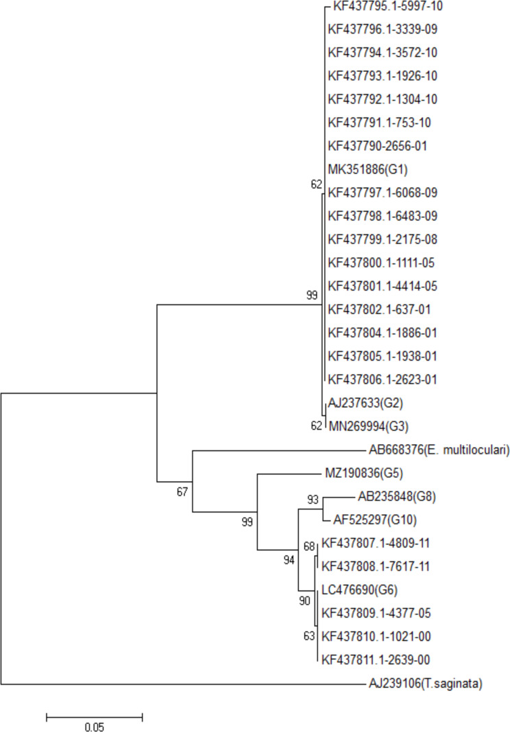
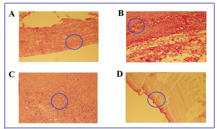
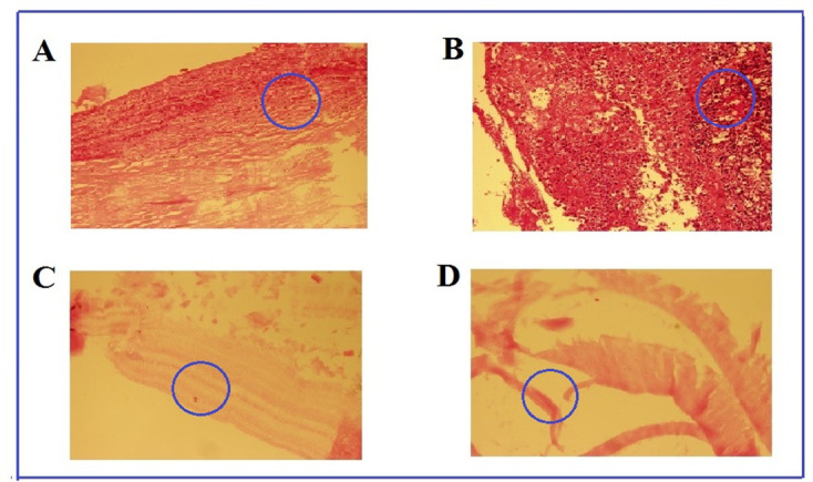
Results: Based on the sequencing results, 16 cases were diagnosed as E. granulosus s.s. (G1-G3 genotype) and 5 cases as E. canadensis (G6 genotype). Five haplotypes of E. granulosus were identified from 21 nad1 sequences. The histopathological alterations in both genotypes showed laminated layer of CE without inflammatory cells. In a few cases of the G6 genotype, neutrophils in the outer cuticular layer with mild vascular and congestion were observed. Cell debris with multiple areas of necrosis, as well as scanty lymphoplasma cells in the outer cuticular layer were observed in G1-G3 genotype cases. So, the histopathological differences between the two genotypes are not noticeable enough to be differentiated by microscopical observations.
Conclusion: E. granulosus s.s. (G1-G3) and E. canadensis (G6 genotype) are prevalent among CE patients. In general, five haplotypes were identified by nad1 genes analysis. The histopathological differences between the two genotypes have not been so big to be differentiated by microscopic observations.
期刊介绍:
Iranian Journal of Parasitology (IJP) is the official publication of Iranian Society of Parasitology (ISP) launched in 2006. The society was inaugurated in 1994 and pursues the improvement of the knowledge on the parasites and parasitic diseases, exchange of scientific knowledge with foreign societies, publicity activities, and consultation on the parasitic diseases, and intimate relationship among society members.
The main aims of the Journal are: contribution to the field of Parasitology, including all aspects of parasites and parasitic diseases (medical and veterinary) and related fields such as Entomology which may be submitted by scientists from Iran and all over the world.
 求助内容:
求助内容: 应助结果提醒方式:


