Clara López-Martínez, Íñigo Aragón-Niño, Alba García-López-Chicharro, Marta María Pampín-Martínez, José Luis Cebrián-Carretero
{"title":"经宫颈入路咽旁间隙多形性腺瘤的计算机辅助切除。病例报告。","authors":"Clara López-Martínez, Íñigo Aragón-Niño, Alba García-López-Chicharro, Marta María Pampín-Martínez, José Luis Cebrián-Carretero","doi":"10.4317/jced.62547","DOIUrl":null,"url":null,"abstract":"<p><strong>Background: </strong>The parapharyngeal space, situated in the neck's lateral part, poses challenges for surgical intervention due to its complex anatomy. Tumors in this space are rare, accounting for 0.5-1% of head and neck tumors, often remaining asymptomatic until reaching substantial sizes.</p><p><strong>Material and methods: </strong>This article outlines the surgical management of such lesions using navigation software, illustrated through a clinical case. A 38-year-old male presented with a mass at the right mandibular angle. Preoperative planning with Brainlab iPlan CMF® software aided in understanding the tumor's relationship with adjacent structures. A transcervical approach facilitated tumor excision without damaging vital anatomical structures. Histopathology confirmed pleomorphic adenoma.</p><p><strong>Results: </strong>The paper underscores the rarity of parapharyngeal tumors, the importance of individualized surgical strategies, and the utility of 3D technology in preoperative planning for enhanced anatomical understanding.</p><p><strong>Conclusions: </strong>The cervical approach remains a satisfactory choice for benign lesions in this challenging space. <b>Key words:</b>Parapharyngeal space, 3D technology, pleomorphic adenoma, transcervical approach, virtual planning.</p>","PeriodicalId":15376,"journal":{"name":"Journal of Clinical and Experimental Dentistry","volume":"17 3","pages":"e350-e353"},"PeriodicalIF":0.0000,"publicationDate":"2025-03-01","publicationTypes":"Journal Article","fieldsOfStudy":null,"isOpenAccess":false,"openAccessPdf":"https://www.ncbi.nlm.nih.gov/pmc/articles/PMC11994214/pdf/","citationCount":"0","resultStr":"{\"title\":\"Computer-Aided excision of Parapharyngeal Space Pleomorphic Adenoma by Transcervical Approach. Case report.\",\"authors\":\"Clara López-Martínez, Íñigo Aragón-Niño, Alba García-López-Chicharro, Marta María Pampín-Martínez, José Luis Cebrián-Carretero\",\"doi\":\"10.4317/jced.62547\",\"DOIUrl\":null,\"url\":null,\"abstract\":\"<p><strong>Background: </strong>The parapharyngeal space, situated in the neck's lateral part, poses challenges for surgical intervention due to its complex anatomy. Tumors in this space are rare, accounting for 0.5-1% of head and neck tumors, often remaining asymptomatic until reaching substantial sizes.</p><p><strong>Material and methods: </strong>This article outlines the surgical management of such lesions using navigation software, illustrated through a clinical case. A 38-year-old male presented with a mass at the right mandibular angle. Preoperative planning with Brainlab iPlan CMF® software aided in understanding the tumor's relationship with adjacent structures. A transcervical approach facilitated tumor excision without damaging vital anatomical structures. Histopathology confirmed pleomorphic adenoma.</p><p><strong>Results: </strong>The paper underscores the rarity of parapharyngeal tumors, the importance of individualized surgical strategies, and the utility of 3D technology in preoperative planning for enhanced anatomical understanding.</p><p><strong>Conclusions: </strong>The cervical approach remains a satisfactory choice for benign lesions in this challenging space. <b>Key words:</b>Parapharyngeal space, 3D technology, pleomorphic adenoma, transcervical approach, virtual planning.</p>\",\"PeriodicalId\":15376,\"journal\":{\"name\":\"Journal of Clinical and Experimental Dentistry\",\"volume\":\"17 3\",\"pages\":\"e350-e353\"},\"PeriodicalIF\":0.0000,\"publicationDate\":\"2025-03-01\",\"publicationTypes\":\"Journal Article\",\"fieldsOfStudy\":null,\"isOpenAccess\":false,\"openAccessPdf\":\"https://www.ncbi.nlm.nih.gov/pmc/articles/PMC11994214/pdf/\",\"citationCount\":\"0\",\"resultStr\":null,\"platform\":\"Semanticscholar\",\"paperid\":null,\"PeriodicalName\":\"Journal of Clinical and Experimental Dentistry\",\"FirstCategoryId\":\"1085\",\"ListUrlMain\":\"https://doi.org/10.4317/jced.62547\",\"RegionNum\":0,\"RegionCategory\":null,\"ArticlePicture\":[],\"TitleCN\":null,\"AbstractTextCN\":null,\"PMCID\":null,\"EPubDate\":\"\",\"PubModel\":\"\",\"JCR\":\"Q2\",\"JCRName\":\"Dentistry\",\"Score\":null,\"Total\":0}","platform":"Semanticscholar","paperid":null,"PeriodicalName":"Journal of Clinical and Experimental Dentistry","FirstCategoryId":"1085","ListUrlMain":"https://doi.org/10.4317/jced.62547","RegionNum":0,"RegionCategory":null,"ArticlePicture":[],"TitleCN":null,"AbstractTextCN":null,"PMCID":null,"EPubDate":"","PubModel":"","JCR":"Q2","JCRName":"Dentistry","Score":null,"Total":0}
Computer-Aided excision of Parapharyngeal Space Pleomorphic Adenoma by Transcervical Approach. Case report.
Background: The parapharyngeal space, situated in the neck's lateral part, poses challenges for surgical intervention due to its complex anatomy. Tumors in this space are rare, accounting for 0.5-1% of head and neck tumors, often remaining asymptomatic until reaching substantial sizes.
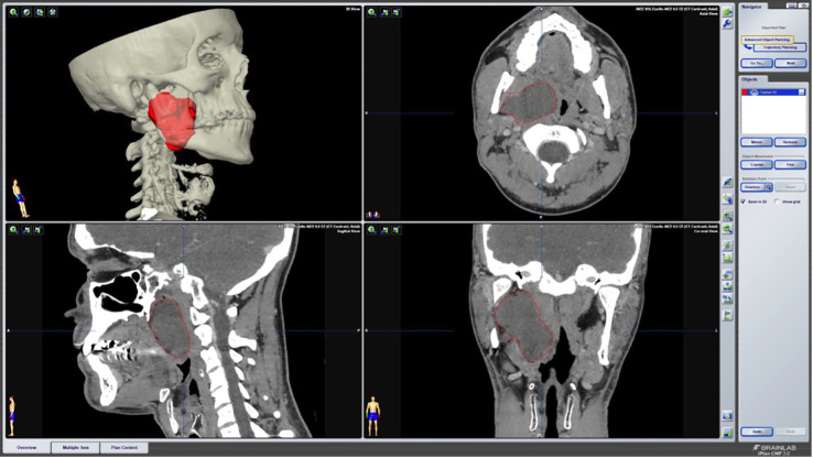
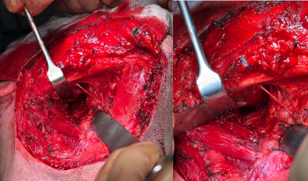
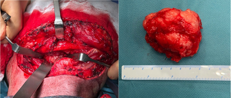
Material and methods: This article outlines the surgical management of such lesions using navigation software, illustrated through a clinical case. A 38-year-old male presented with a mass at the right mandibular angle. Preoperative planning with Brainlab iPlan CMF® software aided in understanding the tumor's relationship with adjacent structures. A transcervical approach facilitated tumor excision without damaging vital anatomical structures. Histopathology confirmed pleomorphic adenoma.
Results: The paper underscores the rarity of parapharyngeal tumors, the importance of individualized surgical strategies, and the utility of 3D technology in preoperative planning for enhanced anatomical understanding.
Conclusions: The cervical approach remains a satisfactory choice for benign lesions in this challenging space. Key words:Parapharyngeal space, 3D technology, pleomorphic adenoma, transcervical approach, virtual planning.
期刊介绍:
Indexed in PUBMED, PubMed Central® (PMC) since 2012 and SCOPUSJournal of Clinical and Experimental Dentistry is an Open Access (free access on-line) - http://www.medicinaoral.com/odo/indice.htm. The aim of the Journal of Clinical and Experimental Dentistry is: - Periodontology - Community and Preventive Dentistry - Esthetic Dentistry - Biomaterials and Bioengineering in Dentistry - Operative Dentistry and Endodontics - Prosthetic Dentistry - Orthodontics - Oral Medicine and Pathology - Odontostomatology for the disabled or special patients - Oral Surgery

 求助内容:
求助内容: 应助结果提醒方式:


