Taylor C Chan, Elisa Heacock, Ashleigh Cournoyer, Koranda Walsh, Amy C Durham
{"title":"猫的透明化胰腺腺癌。","authors":"Taylor C Chan, Elisa Heacock, Ashleigh Cournoyer, Koranda Walsh, Amy C Durham","doi":"10.1177/20551169251325333","DOIUrl":null,"url":null,"abstract":"<p><strong>Case summary: </strong>A 6-year-old female spayed domestic shorthair cat was presented for abdominal distension and weight loss. Abdominal radiographs and ultrasound revealed two cranial abdominal masses and another mass adjacent to the jejunum. Cytologic features of the cranial abdominal masses were consistent with exocrine pancreatic tissue. Four months later, a repeat abdominal ultrasound revealed progressive enlargement of the abdominal masses and medial iliac lymphadenopathy. On exploratory laparotomy, two abdominal masses were associated with the pancreas and incorporated large blood vessels supplying the liver, pancreas and spleen. The masses were non-resectable and incisional biopsies were obtained. The histologic features were diagnostic for a hyalinizing subtype of exocrine pancreatic adenocarcinoma. Chemotherapy was not pursued. Over 28 months after the initial detection of abdominal masses, the cat was still alive and reportedly doing well.</p><p><strong>Relevance and novel information: </strong>To the authors' knowledge, this is the first report of a hyalinizing subtype of pancreatic adenocarcinoma in a cat. This subtype is considered to behave less aggressively in dogs, and this case may support that a similar, more indolent behavior may be seen in cats.</p>","PeriodicalId":36588,"journal":{"name":"Journal of Feline Medicine and Surgery Open Reports","volume":"11 1","pages":"20551169251325333"},"PeriodicalIF":0.7000,"publicationDate":"2025-04-16","publicationTypes":"Journal Article","fieldsOfStudy":null,"isOpenAccess":false,"openAccessPdf":"https://www.ncbi.nlm.nih.gov/pmc/articles/PMC12033424/pdf/","citationCount":"0","resultStr":"{\"title\":\"Hyalinizing pancreatic adenocarcinoma in a cat.\",\"authors\":\"Taylor C Chan, Elisa Heacock, Ashleigh Cournoyer, Koranda Walsh, Amy C Durham\",\"doi\":\"10.1177/20551169251325333\",\"DOIUrl\":null,\"url\":null,\"abstract\":\"<p><strong>Case summary: </strong>A 6-year-old female spayed domestic shorthair cat was presented for abdominal distension and weight loss. Abdominal radiographs and ultrasound revealed two cranial abdominal masses and another mass adjacent to the jejunum. Cytologic features of the cranial abdominal masses were consistent with exocrine pancreatic tissue. Four months later, a repeat abdominal ultrasound revealed progressive enlargement of the abdominal masses and medial iliac lymphadenopathy. On exploratory laparotomy, two abdominal masses were associated with the pancreas and incorporated large blood vessels supplying the liver, pancreas and spleen. The masses were non-resectable and incisional biopsies were obtained. The histologic features were diagnostic for a hyalinizing subtype of exocrine pancreatic adenocarcinoma. Chemotherapy was not pursued. Over 28 months after the initial detection of abdominal masses, the cat was still alive and reportedly doing well.</p><p><strong>Relevance and novel information: </strong>To the authors' knowledge, this is the first report of a hyalinizing subtype of pancreatic adenocarcinoma in a cat. This subtype is considered to behave less aggressively in dogs, and this case may support that a similar, more indolent behavior may be seen in cats.</p>\",\"PeriodicalId\":36588,\"journal\":{\"name\":\"Journal of Feline Medicine and Surgery Open Reports\",\"volume\":\"11 1\",\"pages\":\"20551169251325333\"},\"PeriodicalIF\":0.7000,\"publicationDate\":\"2025-04-16\",\"publicationTypes\":\"Journal Article\",\"fieldsOfStudy\":null,\"isOpenAccess\":false,\"openAccessPdf\":\"https://www.ncbi.nlm.nih.gov/pmc/articles/PMC12033424/pdf/\",\"citationCount\":\"0\",\"resultStr\":null,\"platform\":\"Semanticscholar\",\"paperid\":null,\"PeriodicalName\":\"Journal of Feline Medicine and Surgery Open Reports\",\"FirstCategoryId\":\"1085\",\"ListUrlMain\":\"https://doi.org/10.1177/20551169251325333\",\"RegionNum\":0,\"RegionCategory\":null,\"ArticlePicture\":[],\"TitleCN\":null,\"AbstractTextCN\":null,\"PMCID\":null,\"EPubDate\":\"2025/1/1 0:00:00\",\"PubModel\":\"eCollection\",\"JCR\":\"Q3\",\"JCRName\":\"VETERINARY SCIENCES\",\"Score\":null,\"Total\":0}","platform":"Semanticscholar","paperid":null,"PeriodicalName":"Journal of Feline Medicine and Surgery Open Reports","FirstCategoryId":"1085","ListUrlMain":"https://doi.org/10.1177/20551169251325333","RegionNum":0,"RegionCategory":null,"ArticlePicture":[],"TitleCN":null,"AbstractTextCN":null,"PMCID":null,"EPubDate":"2025/1/1 0:00:00","PubModel":"eCollection","JCR":"Q3","JCRName":"VETERINARY SCIENCES","Score":null,"Total":0}
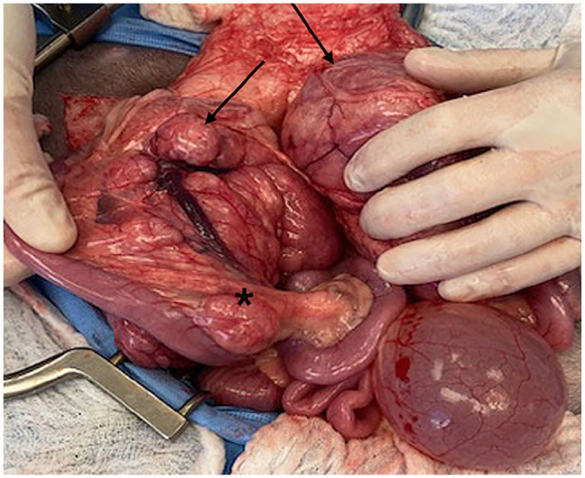
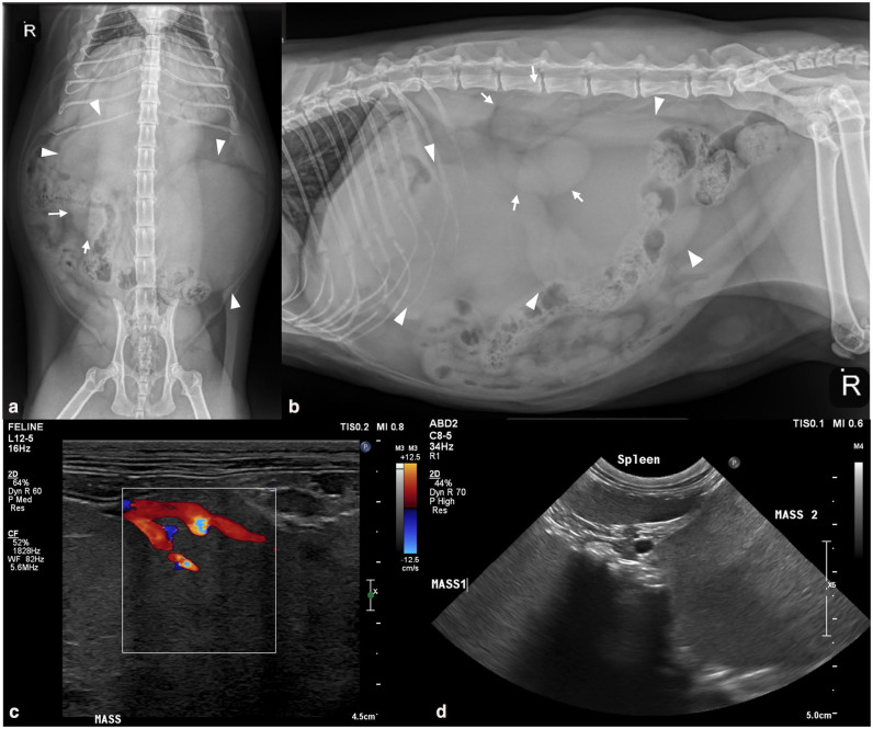
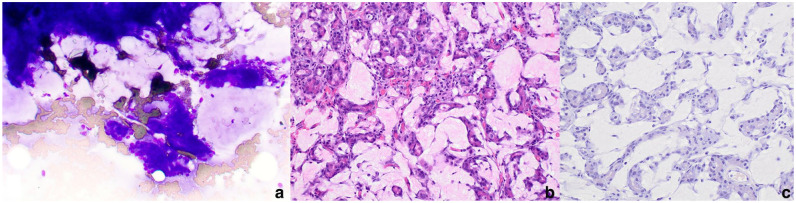
Case summary: A 6-year-old female spayed domestic shorthair cat was presented for abdominal distension and weight loss. Abdominal radiographs and ultrasound revealed two cranial abdominal masses and another mass adjacent to the jejunum. Cytologic features of the cranial abdominal masses were consistent with exocrine pancreatic tissue. Four months later, a repeat abdominal ultrasound revealed progressive enlargement of the abdominal masses and medial iliac lymphadenopathy. On exploratory laparotomy, two abdominal masses were associated with the pancreas and incorporated large blood vessels supplying the liver, pancreas and spleen. The masses were non-resectable and incisional biopsies were obtained. The histologic features were diagnostic for a hyalinizing subtype of exocrine pancreatic adenocarcinoma. Chemotherapy was not pursued. Over 28 months after the initial detection of abdominal masses, the cat was still alive and reportedly doing well.
Relevance and novel information: To the authors' knowledge, this is the first report of a hyalinizing subtype of pancreatic adenocarcinoma in a cat. This subtype is considered to behave less aggressively in dogs, and this case may support that a similar, more indolent behavior may be seen in cats.

 求助内容:
求助内容: 应助结果提醒方式:


