Thiago Paiva Freire, Geraldo Braz Júnior, João Dallyson Sousa de Almeida, José Ribamar Durand Rodrigues Junior
{"title":"基于复合主干和双解码器结构的智能手机手持式检眼镜图像杯盘分割。","authors":"Thiago Paiva Freire, Geraldo Braz Júnior, João Dallyson Sousa de Almeida, José Ribamar Durand Rodrigues Junior","doi":"10.3390/vision9020032","DOIUrl":null,"url":null,"abstract":"<p><p>Glaucoma is a visual disease that affects millions of people, and early diagnosis can prevent total blindness. One way to diagnose the disease is through fundus image examination, which analyzes the optic disc and cup structures. However, screening programs in primary care are costly and unfeasible. Neural network models have been used to segment optic nerve structures, assisting physicians in this task and reducing fatigue. This work presents a methodology to enhance morphological biomarkers of the optic disc and cup in images obtained by a smartphone coupled to an ophthalmoscope through a deep neural network, which combines two backbones and a dual decoder approach to improve the segmentation of these structures, as well as a new way to combine the loss weights in the training process. The models obtained were numerically evaluated through Dice and IoU measures. The dice values obtained in the experiments reached a Dice of 95.92% and 85.30% for the optical disc and cup and an IoU of 92.22% and 75.68% for the optical disc and cup, respectively, in the BrG dataset. These findings indicate promising architectures in the fundus image segmentation task.</p>","PeriodicalId":36586,"journal":{"name":"Vision (Switzerland)","volume":"9 2","pages":""},"PeriodicalIF":1.8000,"publicationDate":"2025-04-11","publicationTypes":"Journal Article","fieldsOfStudy":null,"isOpenAccess":false,"openAccessPdf":"https://www.ncbi.nlm.nih.gov/pmc/articles/PMC12015843/pdf/","citationCount":"0","resultStr":"{\"title\":\"Cup and Disc Segmentation in Smartphone Handheld Ophthalmoscope Images with a Composite Backbone and Double Decoder Architecture.\",\"authors\":\"Thiago Paiva Freire, Geraldo Braz Júnior, João Dallyson Sousa de Almeida, José Ribamar Durand Rodrigues Junior\",\"doi\":\"10.3390/vision9020032\",\"DOIUrl\":null,\"url\":null,\"abstract\":\"<p><p>Glaucoma is a visual disease that affects millions of people, and early diagnosis can prevent total blindness. One way to diagnose the disease is through fundus image examination, which analyzes the optic disc and cup structures. However, screening programs in primary care are costly and unfeasible. Neural network models have been used to segment optic nerve structures, assisting physicians in this task and reducing fatigue. This work presents a methodology to enhance morphological biomarkers of the optic disc and cup in images obtained by a smartphone coupled to an ophthalmoscope through a deep neural network, which combines two backbones and a dual decoder approach to improve the segmentation of these structures, as well as a new way to combine the loss weights in the training process. The models obtained were numerically evaluated through Dice and IoU measures. The dice values obtained in the experiments reached a Dice of 95.92% and 85.30% for the optical disc and cup and an IoU of 92.22% and 75.68% for the optical disc and cup, respectively, in the BrG dataset. These findings indicate promising architectures in the fundus image segmentation task.</p>\",\"PeriodicalId\":36586,\"journal\":{\"name\":\"Vision (Switzerland)\",\"volume\":\"9 2\",\"pages\":\"\"},\"PeriodicalIF\":1.8000,\"publicationDate\":\"2025-04-11\",\"publicationTypes\":\"Journal Article\",\"fieldsOfStudy\":null,\"isOpenAccess\":false,\"openAccessPdf\":\"https://www.ncbi.nlm.nih.gov/pmc/articles/PMC12015843/pdf/\",\"citationCount\":\"0\",\"resultStr\":null,\"platform\":\"Semanticscholar\",\"paperid\":null,\"PeriodicalName\":\"Vision (Switzerland)\",\"FirstCategoryId\":\"1085\",\"ListUrlMain\":\"https://doi.org/10.3390/vision9020032\",\"RegionNum\":0,\"RegionCategory\":null,\"ArticlePicture\":[],\"TitleCN\":null,\"AbstractTextCN\":null,\"PMCID\":null,\"EPubDate\":\"\",\"PubModel\":\"\",\"JCR\":\"Q2\",\"JCRName\":\"Medicine\",\"Score\":null,\"Total\":0}","platform":"Semanticscholar","paperid":null,"PeriodicalName":"Vision (Switzerland)","FirstCategoryId":"1085","ListUrlMain":"https://doi.org/10.3390/vision9020032","RegionNum":0,"RegionCategory":null,"ArticlePicture":[],"TitleCN":null,"AbstractTextCN":null,"PMCID":null,"EPubDate":"","PubModel":"","JCR":"Q2","JCRName":"Medicine","Score":null,"Total":0}
Cup and Disc Segmentation in Smartphone Handheld Ophthalmoscope Images with a Composite Backbone and Double Decoder Architecture.
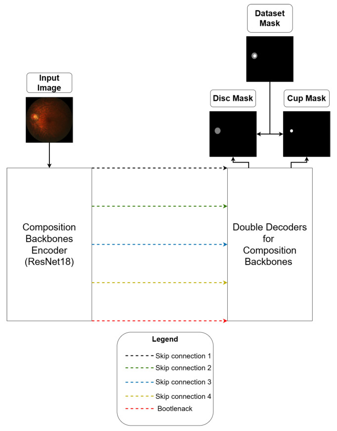
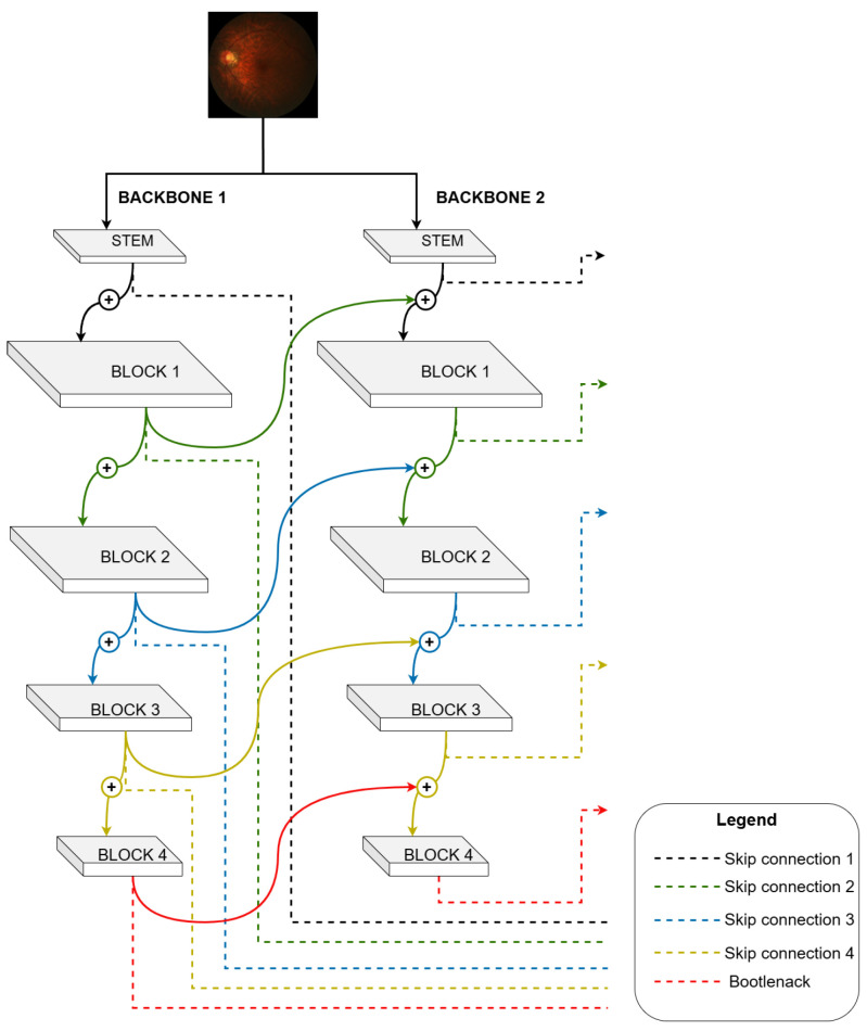
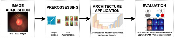
Glaucoma is a visual disease that affects millions of people, and early diagnosis can prevent total blindness. One way to diagnose the disease is through fundus image examination, which analyzes the optic disc and cup structures. However, screening programs in primary care are costly and unfeasible. Neural network models have been used to segment optic nerve structures, assisting physicians in this task and reducing fatigue. This work presents a methodology to enhance morphological biomarkers of the optic disc and cup in images obtained by a smartphone coupled to an ophthalmoscope through a deep neural network, which combines two backbones and a dual decoder approach to improve the segmentation of these structures, as well as a new way to combine the loss weights in the training process. The models obtained were numerically evaluated through Dice and IoU measures. The dice values obtained in the experiments reached a Dice of 95.92% and 85.30% for the optical disc and cup and an IoU of 92.22% and 75.68% for the optical disc and cup, respectively, in the BrG dataset. These findings indicate promising architectures in the fundus image segmentation task.

 求助内容:
求助内容: 应助结果提醒方式:


