{"title":"两种数字制作的冠外修复体:嵌体和全冠:一项前瞻性、交叉弓随机研究,对修复体和临床结果的影响进行了比较评估。","authors":"Sarojini Biswal, Bhupender Kumar Yadav, Abhishek Nagpal, Omkar Krishna Shetty, Pankaj Ritwal, Shalini Kapoor","doi":"10.4103/jips.jips_331_24","DOIUrl":null,"url":null,"abstract":"<p><strong>Aim: </strong>The aim of the study was to investigate and compare the prosthetic parameters, clinical indices, and survival rates of two digitally fabricated extracoronal restorations, namely an onlay and a full crown, at baseline, 1 year, and 2 years.</p><p><strong>Settings and design: </strong>This was a prospective clinical study conducted on endodontically treated posterior teeth.</p><p><strong>Materials and methods: </strong>Endodontically treated posterior teeth (n = 15) present bilaterally in the mandibular arch were selected. Digitally fabricated lithium disilicate onlay and crown were placed on either side of the same arch after randomization. Clinical parameters and prosthetic characteristics (as per the modified United States Public Health Service criteria) were evaluated at baseline, 1-year, and 2-year follow-ups.</p><p><strong>Statistical analysis used: </strong>Data were descriptively examined. The results were evaluated using the Chi-square test and ANOVA. Statistical significance was determined at P < 0.05.</p><p><strong>Results: </strong>Full crowns showed a 100% survival rate, while onlays had an 83.3% survival rate, with a significant difference (P = 0.030, 95% confidence interval: 0.01-0.12). Onlays exhibited significantly better periodontal outcomes, including lower bleeding on probing (P = 0.000), plaque index (P = 0.001), and probing pocket depth (P = 0.000) at 1 and 2 years. Marginal discoloration (20%) and marginal integrity loss (13.3%) were observed in onlays, with significant differences (P = 0.001). Both groups showed no fractures, secondary caries, or significant surface texture changes, and 100% patient satisfaction throughout.</p><p><strong>Conclusion: </strong>In this clinical trial comparing lithium disilicate onlays and crowns for restoring posterior teeth following endodontic treatment, both options demonstrated satisfactory prosthetic parameters during subsequent follow-ups. However, marginal integrity and discoloration were more prevalent in the onlay group. Periodontal examination revealed that onlays exhibited superior periodontal outcomes compared to crowns, with full crowns showing greater periodontal damage at 1-year and 2-year follow-ups.</p>","PeriodicalId":22669,"journal":{"name":"The Journal of Indian Prosthodontic Society","volume":"25 2","pages":"150-162"},"PeriodicalIF":1.0000,"publicationDate":"2025-04-01","publicationTypes":"Journal Article","fieldsOfStudy":null,"isOpenAccess":false,"openAccessPdf":"https://www.ncbi.nlm.nih.gov/pmc/articles/PMC12057828/pdf/","citationCount":"0","resultStr":"{\"title\":\"A comparative evaluation of prosthetic and clinical outcomes influenced by two digitally fabricated extracoronal restorations: An onlay and a full crown: A prospective, cross-arch randomized study.\",\"authors\":\"Sarojini Biswal, Bhupender Kumar Yadav, Abhishek Nagpal, Omkar Krishna Shetty, Pankaj Ritwal, Shalini Kapoor\",\"doi\":\"10.4103/jips.jips_331_24\",\"DOIUrl\":null,\"url\":null,\"abstract\":\"<p><strong>Aim: </strong>The aim of the study was to investigate and compare the prosthetic parameters, clinical indices, and survival rates of two digitally fabricated extracoronal restorations, namely an onlay and a full crown, at baseline, 1 year, and 2 years.</p><p><strong>Settings and design: </strong>This was a prospective clinical study conducted on endodontically treated posterior teeth.</p><p><strong>Materials and methods: </strong>Endodontically treated posterior teeth (n = 15) present bilaterally in the mandibular arch were selected. Digitally fabricated lithium disilicate onlay and crown were placed on either side of the same arch after randomization. Clinical parameters and prosthetic characteristics (as per the modified United States Public Health Service criteria) were evaluated at baseline, 1-year, and 2-year follow-ups.</p><p><strong>Statistical analysis used: </strong>Data were descriptively examined. The results were evaluated using the Chi-square test and ANOVA. Statistical significance was determined at P < 0.05.</p><p><strong>Results: </strong>Full crowns showed a 100% survival rate, while onlays had an 83.3% survival rate, with a significant difference (P = 0.030, 95% confidence interval: 0.01-0.12). Onlays exhibited significantly better periodontal outcomes, including lower bleeding on probing (P = 0.000), plaque index (P = 0.001), and probing pocket depth (P = 0.000) at 1 and 2 years. Marginal discoloration (20%) and marginal integrity loss (13.3%) were observed in onlays, with significant differences (P = 0.001). Both groups showed no fractures, secondary caries, or significant surface texture changes, and 100% patient satisfaction throughout.</p><p><strong>Conclusion: </strong>In this clinical trial comparing lithium disilicate onlays and crowns for restoring posterior teeth following endodontic treatment, both options demonstrated satisfactory prosthetic parameters during subsequent follow-ups. However, marginal integrity and discoloration were more prevalent in the onlay group. Periodontal examination revealed that onlays exhibited superior periodontal outcomes compared to crowns, with full crowns showing greater periodontal damage at 1-year and 2-year follow-ups.</p>\",\"PeriodicalId\":22669,\"journal\":{\"name\":\"The Journal of Indian Prosthodontic Society\",\"volume\":\"25 2\",\"pages\":\"150-162\"},\"PeriodicalIF\":1.0000,\"publicationDate\":\"2025-04-01\",\"publicationTypes\":\"Journal Article\",\"fieldsOfStudy\":null,\"isOpenAccess\":false,\"openAccessPdf\":\"https://www.ncbi.nlm.nih.gov/pmc/articles/PMC12057828/pdf/\",\"citationCount\":\"0\",\"resultStr\":null,\"platform\":\"Semanticscholar\",\"paperid\":null,\"PeriodicalName\":\"The Journal of Indian Prosthodontic Society\",\"FirstCategoryId\":\"1085\",\"ListUrlMain\":\"https://doi.org/10.4103/jips.jips_331_24\",\"RegionNum\":0,\"RegionCategory\":null,\"ArticlePicture\":[],\"TitleCN\":null,\"AbstractTextCN\":null,\"PMCID\":null,\"EPubDate\":\"2025/4/11 0:00:00\",\"PubModel\":\"Epub\",\"JCR\":\"Q3\",\"JCRName\":\"DENTISTRY, ORAL SURGERY & MEDICINE\",\"Score\":null,\"Total\":0}","platform":"Semanticscholar","paperid":null,"PeriodicalName":"The Journal of Indian Prosthodontic Society","FirstCategoryId":"1085","ListUrlMain":"https://doi.org/10.4103/jips.jips_331_24","RegionNum":0,"RegionCategory":null,"ArticlePicture":[],"TitleCN":null,"AbstractTextCN":null,"PMCID":null,"EPubDate":"2025/4/11 0:00:00","PubModel":"Epub","JCR":"Q3","JCRName":"DENTISTRY, ORAL SURGERY & MEDICINE","Score":null,"Total":0}
A comparative evaluation of prosthetic and clinical outcomes influenced by two digitally fabricated extracoronal restorations: An onlay and a full crown: A prospective, cross-arch randomized study.
Aim: The aim of the study was to investigate and compare the prosthetic parameters, clinical indices, and survival rates of two digitally fabricated extracoronal restorations, namely an onlay and a full crown, at baseline, 1 year, and 2 years.
Settings and design: This was a prospective clinical study conducted on endodontically treated posterior teeth.
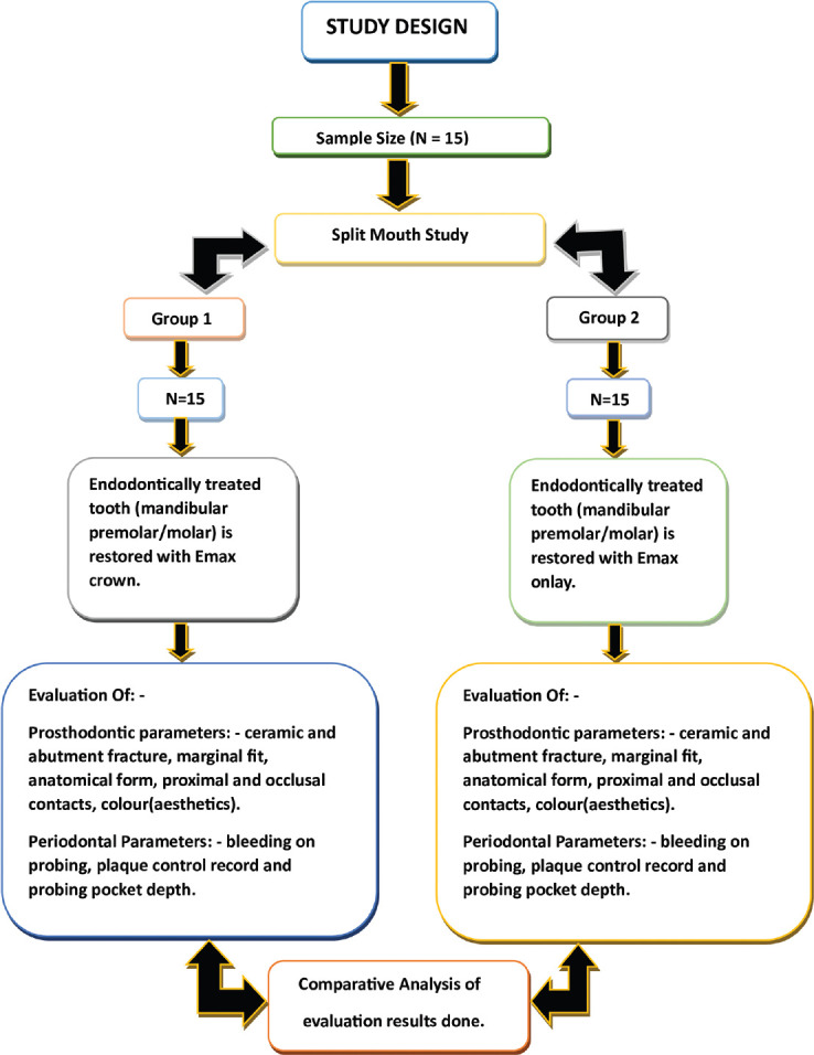
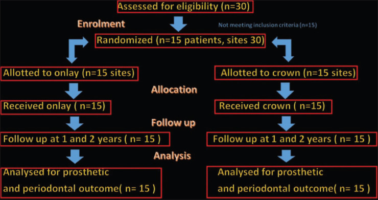
Materials and methods: Endodontically treated posterior teeth (n = 15) present bilaterally in the mandibular arch were selected. Digitally fabricated lithium disilicate onlay and crown were placed on either side of the same arch after randomization. Clinical parameters and prosthetic characteristics (as per the modified United States Public Health Service criteria) were evaluated at baseline, 1-year, and 2-year follow-ups.
Statistical analysis used: Data were descriptively examined. The results were evaluated using the Chi-square test and ANOVA. Statistical significance was determined at P < 0.05.
Results: Full crowns showed a 100% survival rate, while onlays had an 83.3% survival rate, with a significant difference (P = 0.030, 95% confidence interval: 0.01-0.12). Onlays exhibited significantly better periodontal outcomes, including lower bleeding on probing (P = 0.000), plaque index (P = 0.001), and probing pocket depth (P = 0.000) at 1 and 2 years. Marginal discoloration (20%) and marginal integrity loss (13.3%) were observed in onlays, with significant differences (P = 0.001). Both groups showed no fractures, secondary caries, or significant surface texture changes, and 100% patient satisfaction throughout.
Conclusion: In this clinical trial comparing lithium disilicate onlays and crowns for restoring posterior teeth following endodontic treatment, both options demonstrated satisfactory prosthetic parameters during subsequent follow-ups. However, marginal integrity and discoloration were more prevalent in the onlay group. Periodontal examination revealed that onlays exhibited superior periodontal outcomes compared to crowns, with full crowns showing greater periodontal damage at 1-year and 2-year follow-ups.

 求助内容:
求助内容: 应助结果提醒方式:


