Giorgio Tamborrini, Raphael Micheroli, Vincenzo Ricci, Marco Becciolini, Mario Garcia-Pompermayer, Andres Serrano Belmar Gonzalo, Mireille Toranelli, Felix Margenfeld, Magdalena Müller-Gerbl
{"title":"通过组织学和解剖学驱动的高分辨率肌肉骨骼超声增强膝关节成像。","authors":"Giorgio Tamborrini, Raphael Micheroli, Vincenzo Ricci, Marco Becciolini, Mario Garcia-Pompermayer, Andres Serrano Belmar Gonzalo, Mireille Toranelli, Felix Margenfeld, Magdalena Müller-Gerbl","doi":"10.15557/jou.2025.0008","DOIUrl":null,"url":null,"abstract":"<p><strong>Purpose: </strong>To provide an overview of the normal anatomy of the knee using high-resolution ultrasonography.</p><p><strong>Materials and methods: </strong>Normal ultrasound images were obtained from a healthy subject, and corresponding images of human anatomy and histology were acquired from body donors.</p><p><strong>Results: </strong>Several high-resolution ultrasound, anatomical, and histological images were created to illustrate and comprehensively describe the basic standard scans in compliance with international standards. This atlas summarizes a selection of typical normal findings.</p><p><strong>Conclusions: </strong>This overview explains the normal anatomy of the knee as seen by ultrasonography. High-resolution knee musculoskeletal ultrasonography aims to provide an accurate structural evaluation, which requires comprehensive knowledge of sonoanatomy. When used appropriately, contemporary high-resolution musculoskeletal ultrasonography enhances knee imaging by connecting anatomical cross-sections and intricate histology to specific anatomical features.</p>","PeriodicalId":45612,"journal":{"name":"Journal of Ultrasonography","volume":"25 100","pages":"20250008"},"PeriodicalIF":1.5000,"publicationDate":"2025-03-27","publicationTypes":"Journal Article","fieldsOfStudy":null,"isOpenAccess":false,"openAccessPdf":"https://www.ncbi.nlm.nih.gov/pmc/articles/PMC11993308/pdf/","citationCount":"0","resultStr":"{\"title\":\"Enhancing knee imaging via histology and anatomy-driven high-resolution musculoskeletal ultrasound.\",\"authors\":\"Giorgio Tamborrini, Raphael Micheroli, Vincenzo Ricci, Marco Becciolini, Mario Garcia-Pompermayer, Andres Serrano Belmar Gonzalo, Mireille Toranelli, Felix Margenfeld, Magdalena Müller-Gerbl\",\"doi\":\"10.15557/jou.2025.0008\",\"DOIUrl\":null,\"url\":null,\"abstract\":\"<p><strong>Purpose: </strong>To provide an overview of the normal anatomy of the knee using high-resolution ultrasonography.</p><p><strong>Materials and methods: </strong>Normal ultrasound images were obtained from a healthy subject, and corresponding images of human anatomy and histology were acquired from body donors.</p><p><strong>Results: </strong>Several high-resolution ultrasound, anatomical, and histological images were created to illustrate and comprehensively describe the basic standard scans in compliance with international standards. This atlas summarizes a selection of typical normal findings.</p><p><strong>Conclusions: </strong>This overview explains the normal anatomy of the knee as seen by ultrasonography. High-resolution knee musculoskeletal ultrasonography aims to provide an accurate structural evaluation, which requires comprehensive knowledge of sonoanatomy. When used appropriately, contemporary high-resolution musculoskeletal ultrasonography enhances knee imaging by connecting anatomical cross-sections and intricate histology to specific anatomical features.</p>\",\"PeriodicalId\":45612,\"journal\":{\"name\":\"Journal of Ultrasonography\",\"volume\":\"25 100\",\"pages\":\"20250008\"},\"PeriodicalIF\":1.5000,\"publicationDate\":\"2025-03-27\",\"publicationTypes\":\"Journal Article\",\"fieldsOfStudy\":null,\"isOpenAccess\":false,\"openAccessPdf\":\"https://www.ncbi.nlm.nih.gov/pmc/articles/PMC11993308/pdf/\",\"citationCount\":\"0\",\"resultStr\":null,\"platform\":\"Semanticscholar\",\"paperid\":null,\"PeriodicalName\":\"Journal of Ultrasonography\",\"FirstCategoryId\":\"1085\",\"ListUrlMain\":\"https://doi.org/10.15557/jou.2025.0008\",\"RegionNum\":0,\"RegionCategory\":null,\"ArticlePicture\":[],\"TitleCN\":null,\"AbstractTextCN\":null,\"PMCID\":null,\"EPubDate\":\"2025/1/1 0:00:00\",\"PubModel\":\"eCollection\",\"JCR\":\"Q3\",\"JCRName\":\"RADIOLOGY, NUCLEAR MEDICINE & MEDICAL IMAGING\",\"Score\":null,\"Total\":0}","platform":"Semanticscholar","paperid":null,"PeriodicalName":"Journal of Ultrasonography","FirstCategoryId":"1085","ListUrlMain":"https://doi.org/10.15557/jou.2025.0008","RegionNum":0,"RegionCategory":null,"ArticlePicture":[],"TitleCN":null,"AbstractTextCN":null,"PMCID":null,"EPubDate":"2025/1/1 0:00:00","PubModel":"eCollection","JCR":"Q3","JCRName":"RADIOLOGY, NUCLEAR MEDICINE & MEDICAL IMAGING","Score":null,"Total":0}
Enhancing knee imaging via histology and anatomy-driven high-resolution musculoskeletal ultrasound.
Purpose: To provide an overview of the normal anatomy of the knee using high-resolution ultrasonography.
Materials and methods: Normal ultrasound images were obtained from a healthy subject, and corresponding images of human anatomy and histology were acquired from body donors.
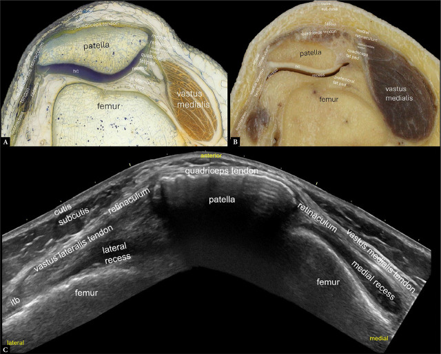
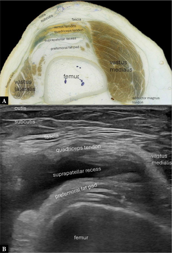
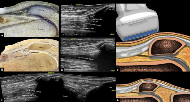
Results: Several high-resolution ultrasound, anatomical, and histological images were created to illustrate and comprehensively describe the basic standard scans in compliance with international standards. This atlas summarizes a selection of typical normal findings.
Conclusions: This overview explains the normal anatomy of the knee as seen by ultrasonography. High-resolution knee musculoskeletal ultrasonography aims to provide an accurate structural evaluation, which requires comprehensive knowledge of sonoanatomy. When used appropriately, contemporary high-resolution musculoskeletal ultrasonography enhances knee imaging by connecting anatomical cross-sections and intricate histology to specific anatomical features.

 求助内容:
求助内容: 应助结果提醒方式:


