V R Arun Kumar, Shivasakthy Manivasakan, K Prabhu, David Livingstone, J Shanti Swarup
{"title":"上颌窦提升后种植体稳定钢板对种植体周围应力分布的影响:一项有限元研究。","authors":"V R Arun Kumar, Shivasakthy Manivasakan, K Prabhu, David Livingstone, J Shanti Swarup","doi":"10.4103/jips.jips_409_24","DOIUrl":null,"url":null,"abstract":"<p><strong>Aim: </strong>To assess the effectiveness of this Implant T Stabilizing plate in distributing the masticatory stress around the implant placed immediately after maxillary sinus lifting in cases with reduced remaining bone height (3 mm).</p><p><strong>Settings and design: </strong>In vitro experimental study.</p><p><strong>Materials and methods: </strong>Two 3D finite element maxillary models with implant placement in 16 region were designed, one without an implant T stabilizing plate and another with an implant T stabilizing plate. Interim removable partial denture with soft liner replacing 16 and 17 was also designed for both models over which the masticatory force of 12 kg was applied. The maximum principal stress generated around the implant and surrounding bone in both models was measured.</p><p><strong>Statistical analysis used: </strong>Statistical analysis was done by using MedCalc stratistical software - version 23.1.7 in which 'Receiver Operating Characteristic Curve' analysis was performed to check for the sensitivity and specificity of the material used.</p><p><strong>Results: </strong>The maximum principal stress around the implant and the cortical bone in working model 1 (without the ITS plate) was 5.2972 and 5.2387 mega pascal respectively. The maximum principal stress around the implant and the cortical bone in working model 2 (with ITS plate) was 1.1663 and 4.5568 mega pascal respectively. In the working model 2, the Implant T stabilizing plate absorbs maximum stress of 10.022 mega pascal.</p><p><strong>Conclusion: </strong>The implant T stabilizing (ITS) plate to the implant placed immediately after maxillary sinus lifting absorbs the maximum stress there by reducing the stress around the implant and surrounding bone even in cases of minimal remaining bone height.</p>","PeriodicalId":22669,"journal":{"name":"The Journal of Indian Prosthodontic Society","volume":"25 2","pages":"138-143"},"PeriodicalIF":1.0000,"publicationDate":"2025-04-01","publicationTypes":"Journal Article","fieldsOfStudy":null,"isOpenAccess":false,"openAccessPdf":"https://www.ncbi.nlm.nih.gov/pmc/articles/PMC12057825/pdf/","citationCount":"0","resultStr":"{\"title\":\"Effectiveness of the implant stabilizing plate on the stress distribution around the implant placed immediately after maxillary sinus lifting: A finite element study.\",\"authors\":\"V R Arun Kumar, Shivasakthy Manivasakan, K Prabhu, David Livingstone, J Shanti Swarup\",\"doi\":\"10.4103/jips.jips_409_24\",\"DOIUrl\":null,\"url\":null,\"abstract\":\"<p><strong>Aim: </strong>To assess the effectiveness of this Implant T Stabilizing plate in distributing the masticatory stress around the implant placed immediately after maxillary sinus lifting in cases with reduced remaining bone height (3 mm).</p><p><strong>Settings and design: </strong>In vitro experimental study.</p><p><strong>Materials and methods: </strong>Two 3D finite element maxillary models with implant placement in 16 region were designed, one without an implant T stabilizing plate and another with an implant T stabilizing plate. Interim removable partial denture with soft liner replacing 16 and 17 was also designed for both models over which the masticatory force of 12 kg was applied. The maximum principal stress generated around the implant and surrounding bone in both models was measured.</p><p><strong>Statistical analysis used: </strong>Statistical analysis was done by using MedCalc stratistical software - version 23.1.7 in which 'Receiver Operating Characteristic Curve' analysis was performed to check for the sensitivity and specificity of the material used.</p><p><strong>Results: </strong>The maximum principal stress around the implant and the cortical bone in working model 1 (without the ITS plate) was 5.2972 and 5.2387 mega pascal respectively. The maximum principal stress around the implant and the cortical bone in working model 2 (with ITS plate) was 1.1663 and 4.5568 mega pascal respectively. In the working model 2, the Implant T stabilizing plate absorbs maximum stress of 10.022 mega pascal.</p><p><strong>Conclusion: </strong>The implant T stabilizing (ITS) plate to the implant placed immediately after maxillary sinus lifting absorbs the maximum stress there by reducing the stress around the implant and surrounding bone even in cases of minimal remaining bone height.</p>\",\"PeriodicalId\":22669,\"journal\":{\"name\":\"The Journal of Indian Prosthodontic Society\",\"volume\":\"25 2\",\"pages\":\"138-143\"},\"PeriodicalIF\":1.0000,\"publicationDate\":\"2025-04-01\",\"publicationTypes\":\"Journal Article\",\"fieldsOfStudy\":null,\"isOpenAccess\":false,\"openAccessPdf\":\"https://www.ncbi.nlm.nih.gov/pmc/articles/PMC12057825/pdf/\",\"citationCount\":\"0\",\"resultStr\":null,\"platform\":\"Semanticscholar\",\"paperid\":null,\"PeriodicalName\":\"The Journal of Indian Prosthodontic Society\",\"FirstCategoryId\":\"1085\",\"ListUrlMain\":\"https://doi.org/10.4103/jips.jips_409_24\",\"RegionNum\":0,\"RegionCategory\":null,\"ArticlePicture\":[],\"TitleCN\":null,\"AbstractTextCN\":null,\"PMCID\":null,\"EPubDate\":\"2025/4/11 0:00:00\",\"PubModel\":\"Epub\",\"JCR\":\"Q3\",\"JCRName\":\"DENTISTRY, ORAL SURGERY & MEDICINE\",\"Score\":null,\"Total\":0}","platform":"Semanticscholar","paperid":null,"PeriodicalName":"The Journal of Indian Prosthodontic Society","FirstCategoryId":"1085","ListUrlMain":"https://doi.org/10.4103/jips.jips_409_24","RegionNum":0,"RegionCategory":null,"ArticlePicture":[],"TitleCN":null,"AbstractTextCN":null,"PMCID":null,"EPubDate":"2025/4/11 0:00:00","PubModel":"Epub","JCR":"Q3","JCRName":"DENTISTRY, ORAL SURGERY & MEDICINE","Score":null,"Total":0}
Effectiveness of the implant stabilizing plate on the stress distribution around the implant placed immediately after maxillary sinus lifting: A finite element study.
Aim: To assess the effectiveness of this Implant T Stabilizing plate in distributing the masticatory stress around the implant placed immediately after maxillary sinus lifting in cases with reduced remaining bone height (3 mm).
Settings and design: In vitro experimental study.
Materials and methods: Two 3D finite element maxillary models with implant placement in 16 region were designed, one without an implant T stabilizing plate and another with an implant T stabilizing plate. Interim removable partial denture with soft liner replacing 16 and 17 was also designed for both models over which the masticatory force of 12 kg was applied. The maximum principal stress generated around the implant and surrounding bone in both models was measured.
Statistical analysis used: Statistical analysis was done by using MedCalc stratistical software - version 23.1.7 in which 'Receiver Operating Characteristic Curve' analysis was performed to check for the sensitivity and specificity of the material used.
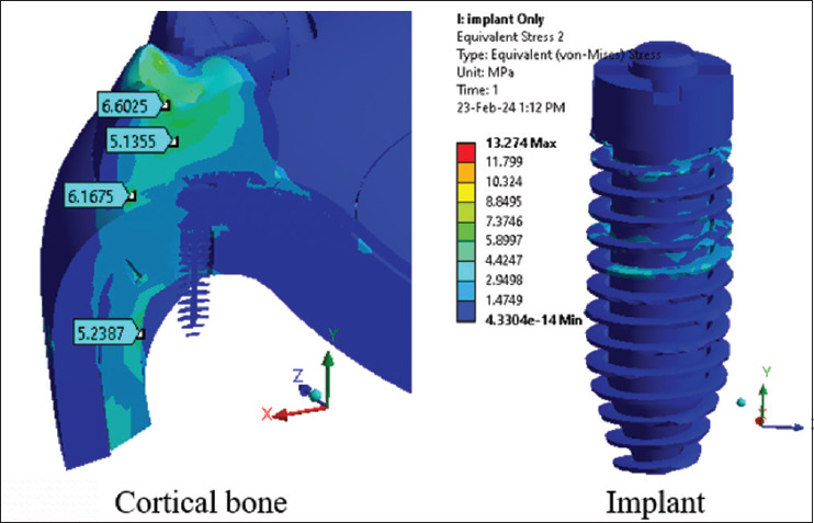
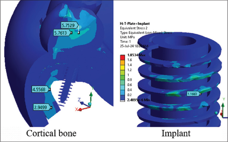
Results: The maximum principal stress around the implant and the cortical bone in working model 1 (without the ITS plate) was 5.2972 and 5.2387 mega pascal respectively. The maximum principal stress around the implant and the cortical bone in working model 2 (with ITS plate) was 1.1663 and 4.5568 mega pascal respectively. In the working model 2, the Implant T stabilizing plate absorbs maximum stress of 10.022 mega pascal.
Conclusion: The implant T stabilizing (ITS) plate to the implant placed immediately after maxillary sinus lifting absorbs the maximum stress there by reducing the stress around the implant and surrounding bone even in cases of minimal remaining bone height.

 求助内容:
求助内容: 应助结果提醒方式:


