{"title":"在离靶不同距离的发光二极管照射下成骨潜能的比较评价:一项体外研究。","authors":"Divyabharathi Selvam, Venkat Rengasamy","doi":"10.4103/jips.jips_488_24","DOIUrl":null,"url":null,"abstract":"<p><strong>Aim: </strong>To evaluate and compare the osteogenic effects of LED irradiation at varying distances using MG-63 osteoblast-like cells cultured on titanium discs.</p><p><strong>Settings and design: </strong>This in vitro experimental study involved human osteosarcoma (MG-63) cells cultured on titanium discs and subjected to LED irradiation at different distances, compared to a non-irradiated control group.</p><p><strong>Materials and methods: </strong>Forty-two titanium discs were divided into three groups: Control (no irradiation), LED Group 1 (10 mm distance), and LED Group 2 (20 mm distance). All discs were seeded with MG-63 cells and pre-cultured. Experimental groups received a single 2-minute exposure to 660 nm red LED light, while controls received no exposure. Cell viability was measured at 24 and 48 hours using the MTT assay. Cell attachment, growth, and proliferation were assessed at 72, 96, and 120 hours.</p><p><strong>Statistical analysis used: </strong>Data were analyzed using SPSS v28.0. Two-way ANOVA followed by Tukey's post-hoc test was applied to identify statistically significant differences among groups across time points. A p-value < 0.05 was considered significant.</p><p><strong>Results: </strong>Both LED-irradiated groups showed significantly enhanced osteogenic activity compared to controls (p < 0.05). Group 1 (10 mm) exhibited the highest cell viability, attachment, and proliferation. Group 2 (20 mm) showed moderate improvement but was inferior to Group 1, indicating distance-dependent effects.</p><p><strong>Conclusion: </strong>LED therapy enhances osteogenesis on titanium surfaces, with the greatest efficacy observed at a 10 mm irradiation distance. These findings support the use of optimized LED therapy to improve bone healing and implant integration.</p>","PeriodicalId":22669,"journal":{"name":"The Journal of Indian Prosthodontic Society","volume":"25 2","pages":"169-175"},"PeriodicalIF":1.0000,"publicationDate":"2025-04-01","publicationTypes":"Journal Article","fieldsOfStudy":null,"isOpenAccess":false,"openAccessPdf":"https://www.ncbi.nlm.nih.gov/pmc/articles/PMC12057822/pdf/","citationCount":"0","resultStr":"{\"title\":\"Comparative evaluation of osteogenic potential of light-emitting diode irradiation at varying distances from the target: An in vitro study.\",\"authors\":\"Divyabharathi Selvam, Venkat Rengasamy\",\"doi\":\"10.4103/jips.jips_488_24\",\"DOIUrl\":null,\"url\":null,\"abstract\":\"<p><strong>Aim: </strong>To evaluate and compare the osteogenic effects of LED irradiation at varying distances using MG-63 osteoblast-like cells cultured on titanium discs.</p><p><strong>Settings and design: </strong>This in vitro experimental study involved human osteosarcoma (MG-63) cells cultured on titanium discs and subjected to LED irradiation at different distances, compared to a non-irradiated control group.</p><p><strong>Materials and methods: </strong>Forty-two titanium discs were divided into three groups: Control (no irradiation), LED Group 1 (10 mm distance), and LED Group 2 (20 mm distance). All discs were seeded with MG-63 cells and pre-cultured. Experimental groups received a single 2-minute exposure to 660 nm red LED light, while controls received no exposure. Cell viability was measured at 24 and 48 hours using the MTT assay. Cell attachment, growth, and proliferation were assessed at 72, 96, and 120 hours.</p><p><strong>Statistical analysis used: </strong>Data were analyzed using SPSS v28.0. Two-way ANOVA followed by Tukey's post-hoc test was applied to identify statistically significant differences among groups across time points. A p-value < 0.05 was considered significant.</p><p><strong>Results: </strong>Both LED-irradiated groups showed significantly enhanced osteogenic activity compared to controls (p < 0.05). Group 1 (10 mm) exhibited the highest cell viability, attachment, and proliferation. Group 2 (20 mm) showed moderate improvement but was inferior to Group 1, indicating distance-dependent effects.</p><p><strong>Conclusion: </strong>LED therapy enhances osteogenesis on titanium surfaces, with the greatest efficacy observed at a 10 mm irradiation distance. These findings support the use of optimized LED therapy to improve bone healing and implant integration.</p>\",\"PeriodicalId\":22669,\"journal\":{\"name\":\"The Journal of Indian Prosthodontic Society\",\"volume\":\"25 2\",\"pages\":\"169-175\"},\"PeriodicalIF\":1.0000,\"publicationDate\":\"2025-04-01\",\"publicationTypes\":\"Journal Article\",\"fieldsOfStudy\":null,\"isOpenAccess\":false,\"openAccessPdf\":\"https://www.ncbi.nlm.nih.gov/pmc/articles/PMC12057822/pdf/\",\"citationCount\":\"0\",\"resultStr\":null,\"platform\":\"Semanticscholar\",\"paperid\":null,\"PeriodicalName\":\"The Journal of Indian Prosthodontic Society\",\"FirstCategoryId\":\"1085\",\"ListUrlMain\":\"https://doi.org/10.4103/jips.jips_488_24\",\"RegionNum\":0,\"RegionCategory\":null,\"ArticlePicture\":[],\"TitleCN\":null,\"AbstractTextCN\":null,\"PMCID\":null,\"EPubDate\":\"2025/4/11 0:00:00\",\"PubModel\":\"Epub\",\"JCR\":\"Q3\",\"JCRName\":\"DENTISTRY, ORAL SURGERY & MEDICINE\",\"Score\":null,\"Total\":0}","platform":"Semanticscholar","paperid":null,"PeriodicalName":"The Journal of Indian Prosthodontic Society","FirstCategoryId":"1085","ListUrlMain":"https://doi.org/10.4103/jips.jips_488_24","RegionNum":0,"RegionCategory":null,"ArticlePicture":[],"TitleCN":null,"AbstractTextCN":null,"PMCID":null,"EPubDate":"2025/4/11 0:00:00","PubModel":"Epub","JCR":"Q3","JCRName":"DENTISTRY, ORAL SURGERY & MEDICINE","Score":null,"Total":0}
Comparative evaluation of osteogenic potential of light-emitting diode irradiation at varying distances from the target: An in vitro study.
Aim: To evaluate and compare the osteogenic effects of LED irradiation at varying distances using MG-63 osteoblast-like cells cultured on titanium discs.
Settings and design: This in vitro experimental study involved human osteosarcoma (MG-63) cells cultured on titanium discs and subjected to LED irradiation at different distances, compared to a non-irradiated control group.
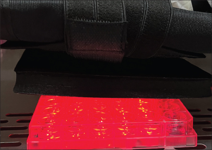
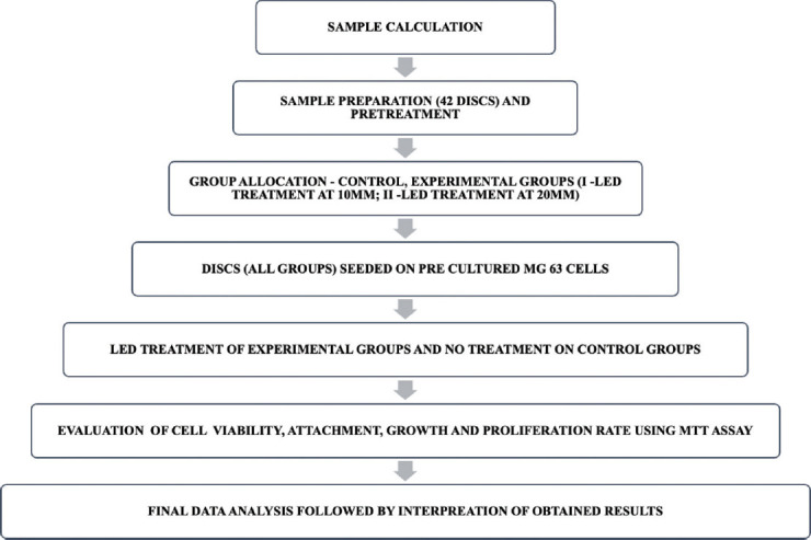
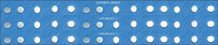
Materials and methods: Forty-two titanium discs were divided into three groups: Control (no irradiation), LED Group 1 (10 mm distance), and LED Group 2 (20 mm distance). All discs were seeded with MG-63 cells and pre-cultured. Experimental groups received a single 2-minute exposure to 660 nm red LED light, while controls received no exposure. Cell viability was measured at 24 and 48 hours using the MTT assay. Cell attachment, growth, and proliferation were assessed at 72, 96, and 120 hours.
Statistical analysis used: Data were analyzed using SPSS v28.0. Two-way ANOVA followed by Tukey's post-hoc test was applied to identify statistically significant differences among groups across time points. A p-value < 0.05 was considered significant.
Results: Both LED-irradiated groups showed significantly enhanced osteogenic activity compared to controls (p < 0.05). Group 1 (10 mm) exhibited the highest cell viability, attachment, and proliferation. Group 2 (20 mm) showed moderate improvement but was inferior to Group 1, indicating distance-dependent effects.
Conclusion: LED therapy enhances osteogenesis on titanium surfaces, with the greatest efficacy observed at a 10 mm irradiation distance. These findings support the use of optimized LED therapy to improve bone healing and implant integration.

 求助内容:
求助内容: 应助结果提醒方式:


