Jun Ma, Junfang Zhang, Mengjia Tan, Min Ji, Jianfeng Yu, Huaijin Guan
{"title":"糖尿病前期角膜神经结构和功能的改变。","authors":"Jun Ma, Junfang Zhang, Mengjia Tan, Min Ji, Jianfeng Yu, Huaijin Guan","doi":"10.1155/jdr/4586856","DOIUrl":null,"url":null,"abstract":"<p><p><b>Background:</b> Although prediabetes increases the risk of developing diabetes, its role in neuropathy remains unclear. We aim to assess alterations in the corneal nerve structure and function in prediabetes and risk factors for corneal nerve loss. <b>Methods:</b> An examination of IVCM and corneal sensitivity was conducted on a cohort of 75 participants, comprising 23 controls, 32 prediabetes, and 20 Type 2 diabetes. Semiautomatic analysis was employed to quantify the corneal nerve fiber length (CNFL), corneal nerve fiber density (CNFD), and dendritic cell (DC) density. <b>Results:</b> CNFL and CNFD were lower in prediabetes and Type 2 diabetes than in the controls, and they were associated with DC density. CNFL and CNFD were lower in Type 2 diabetes than in prediabetes. DC density was higher in prediabetes and Type 2 diabetes than in controls. However, there were no differences in corneal sensitivity between controls and prediabetes. Multivariable regression analysis demonstrated an association between reduced CNFL and age, BMI, fasting plasma glucose (FPG), and uric acid (UA) levels in prediabetes. In Type 2 diabetes, age, HbA1c, blood urea nitrogen (BUN), creatinine (Cr), and triglyceride levels exhibited associations with reduced CNFL. <b>Conclusions:</b> Corneal nerve damage was detected in prediabetes using IVCM. The patients with prediabetes showed signs of nerve structure damage, and the corneal nerve structure damage occurred before the nerve function changes. Immune cells also participate in the occurrence and development of DCN and are related to the corneal neuropathy. Understanding the corneal nerve fiber condition through IVCM may prove crucial in monitoring prediabetic neuropathy.</p>","PeriodicalId":15576,"journal":{"name":"Journal of Diabetes Research","volume":"2025 ","pages":"4586856"},"PeriodicalIF":3.4000,"publicationDate":"2025-04-12","publicationTypes":"Journal Article","fieldsOfStudy":null,"isOpenAccess":false,"openAccessPdf":"https://www.ncbi.nlm.nih.gov/pmc/articles/PMC12009175/pdf/","citationCount":"0","resultStr":"{\"title\":\"Alterations in Corneal Nerve Structure and Function in Prediabetes.\",\"authors\":\"Jun Ma, Junfang Zhang, Mengjia Tan, Min Ji, Jianfeng Yu, Huaijin Guan\",\"doi\":\"10.1155/jdr/4586856\",\"DOIUrl\":null,\"url\":null,\"abstract\":\"<p><p><b>Background:</b> Although prediabetes increases the risk of developing diabetes, its role in neuropathy remains unclear. We aim to assess alterations in the corneal nerve structure and function in prediabetes and risk factors for corneal nerve loss. <b>Methods:</b> An examination of IVCM and corneal sensitivity was conducted on a cohort of 75 participants, comprising 23 controls, 32 prediabetes, and 20 Type 2 diabetes. Semiautomatic analysis was employed to quantify the corneal nerve fiber length (CNFL), corneal nerve fiber density (CNFD), and dendritic cell (DC) density. <b>Results:</b> CNFL and CNFD were lower in prediabetes and Type 2 diabetes than in the controls, and they were associated with DC density. CNFL and CNFD were lower in Type 2 diabetes than in prediabetes. DC density was higher in prediabetes and Type 2 diabetes than in controls. However, there were no differences in corneal sensitivity between controls and prediabetes. Multivariable regression analysis demonstrated an association between reduced CNFL and age, BMI, fasting plasma glucose (FPG), and uric acid (UA) levels in prediabetes. In Type 2 diabetes, age, HbA1c, blood urea nitrogen (BUN), creatinine (Cr), and triglyceride levels exhibited associations with reduced CNFL. <b>Conclusions:</b> Corneal nerve damage was detected in prediabetes using IVCM. The patients with prediabetes showed signs of nerve structure damage, and the corneal nerve structure damage occurred before the nerve function changes. Immune cells also participate in the occurrence and development of DCN and are related to the corneal neuropathy. Understanding the corneal nerve fiber condition through IVCM may prove crucial in monitoring prediabetic neuropathy.</p>\",\"PeriodicalId\":15576,\"journal\":{\"name\":\"Journal of Diabetes Research\",\"volume\":\"2025 \",\"pages\":\"4586856\"},\"PeriodicalIF\":3.4000,\"publicationDate\":\"2025-04-12\",\"publicationTypes\":\"Journal Article\",\"fieldsOfStudy\":null,\"isOpenAccess\":false,\"openAccessPdf\":\"https://www.ncbi.nlm.nih.gov/pmc/articles/PMC12009175/pdf/\",\"citationCount\":\"0\",\"resultStr\":null,\"platform\":\"Semanticscholar\",\"paperid\":null,\"PeriodicalName\":\"Journal of Diabetes Research\",\"FirstCategoryId\":\"3\",\"ListUrlMain\":\"https://doi.org/10.1155/jdr/4586856\",\"RegionNum\":3,\"RegionCategory\":\"医学\",\"ArticlePicture\":[],\"TitleCN\":null,\"AbstractTextCN\":null,\"PMCID\":null,\"EPubDate\":\"2025/1/1 0:00:00\",\"PubModel\":\"eCollection\",\"JCR\":\"Q2\",\"JCRName\":\"ENDOCRINOLOGY & METABOLISM\",\"Score\":null,\"Total\":0}","platform":"Semanticscholar","paperid":null,"PeriodicalName":"Journal of Diabetes Research","FirstCategoryId":"3","ListUrlMain":"https://doi.org/10.1155/jdr/4586856","RegionNum":3,"RegionCategory":"医学","ArticlePicture":[],"TitleCN":null,"AbstractTextCN":null,"PMCID":null,"EPubDate":"2025/1/1 0:00:00","PubModel":"eCollection","JCR":"Q2","JCRName":"ENDOCRINOLOGY & METABOLISM","Score":null,"Total":0}
Alterations in Corneal Nerve Structure and Function in Prediabetes.
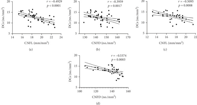
Background: Although prediabetes increases the risk of developing diabetes, its role in neuropathy remains unclear. We aim to assess alterations in the corneal nerve structure and function in prediabetes and risk factors for corneal nerve loss. Methods: An examination of IVCM and corneal sensitivity was conducted on a cohort of 75 participants, comprising 23 controls, 32 prediabetes, and 20 Type 2 diabetes. Semiautomatic analysis was employed to quantify the corneal nerve fiber length (CNFL), corneal nerve fiber density (CNFD), and dendritic cell (DC) density. Results: CNFL and CNFD were lower in prediabetes and Type 2 diabetes than in the controls, and they were associated with DC density. CNFL and CNFD were lower in Type 2 diabetes than in prediabetes. DC density was higher in prediabetes and Type 2 diabetes than in controls. However, there were no differences in corneal sensitivity between controls and prediabetes. Multivariable regression analysis demonstrated an association between reduced CNFL and age, BMI, fasting plasma glucose (FPG), and uric acid (UA) levels in prediabetes. In Type 2 diabetes, age, HbA1c, blood urea nitrogen (BUN), creatinine (Cr), and triglyceride levels exhibited associations with reduced CNFL. Conclusions: Corneal nerve damage was detected in prediabetes using IVCM. The patients with prediabetes showed signs of nerve structure damage, and the corneal nerve structure damage occurred before the nerve function changes. Immune cells also participate in the occurrence and development of DCN and are related to the corneal neuropathy. Understanding the corneal nerve fiber condition through IVCM may prove crucial in monitoring prediabetic neuropathy.
期刊介绍:
Journal of Diabetes Research is a peer-reviewed, Open Access journal that publishes research articles, review articles, and clinical studies related to type 1 and type 2 diabetes. The journal welcomes submissions focusing on the epidemiology, etiology, pathogenesis, management, and prevention of diabetes, as well as associated complications, such as diabetic retinopathy, neuropathy and nephropathy.
 求助内容:
求助内容: 应助结果提醒方式:


