III类倾向患者因生牙和间隙封闭而拔除第11和21牙1例。
摘要
牙齿长牙是一种牙齿现象,单个牙芽试图分裂成两个,导致形成一个结构,看起来像两个牙齿,但起源于同一个毛囊。这种部分分离通常在临床上表现为沟或凹陷,表明存在两颗不同的牙齿(Rajeswari M, Ananthalakshmi R. 2011)。发病-病例报告和复查。印度多学科牙科杂志)。分化和融合的区别在治疗计划中起着重要的作用。如果牙齿数量少一颗,则牙齿融合而不是再生。此外,文献中假设新生牙有一个根管,融合牙有两个独立的根管(Mahendra et al. In Case Rep Dent. 2014:425343, 2014;Duncan and Helpin In Oral surgery Oral Med Oral Pathol 64:82- 87,1987)。牙齿的长出是相对罕见的,主要发生在上颌的额部。单侧牙长出的患病率在初级牙列中为0.01 - 0.04%,在恒牙列中为0.05% (Duncan and Helpin in Oral surgery Oral Med Oral Pathol 64:82- 87,1987)。发芽管理通常需要多学科的方法,涉及几个步骤(Rajeswari M, Ananthalakshmi R. 2011)。发病-病例报告和复查。印度多学科牙科杂志)。然后,正畸医生会对患者进行全面的医疗、牙科和家族史调查,并进行临床和放射检查以确认诊断。治疗选择包括用合适的材料重塑和修复牙齿,进行根管治疗,然后缩小中远端宽度和冠修复,如果牙齿不适合根管治疗,拔牙,然后关闭正畸间隙,或在需要时使用固定或可移动的假体,移植多余的牙齿以取代缺失的牙齿。这个病例报告了一个病人的牙齿长出11,21,并有III级生长的趋势。在这个病例中,畸形的前牙被拔除,并使用骨骼锚固来关闭间隙。缺失中切牙的患者通常需要复杂的跨学科治疗,无论是选择假牙支持修复缺失的前牙,单种植体还是正畸间隙关闭。理想情况下,每种选择都应该满足个人的审美问题、功能要求和牙周组织健康,不仅在治疗结束时,而且在长期内(Marco in Sem orthodon 26:1, 2020;Rosa M, Zachrisson BU。上颌侧切牙缺失患者空间闭合与口腔美容的整合。中华口腔外科杂志;2007;41 (9);Czochrowska定位如何,Skaare,学士, steven nik A, Zachrisson, b.u。缺失上颌中切牙的正畸间隙封闭的疗效[j]。如果选择关闭间隙,选择正确的正畸矫治器和支具是很重要的,特别是对于上颌矢状缺损的III类患者。
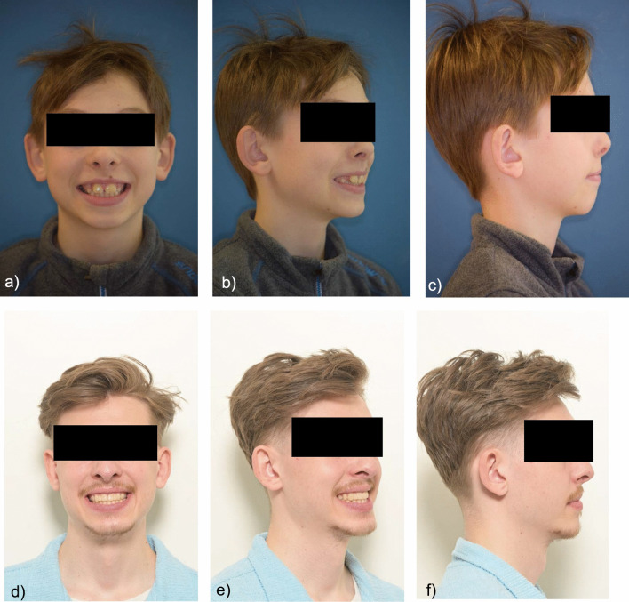
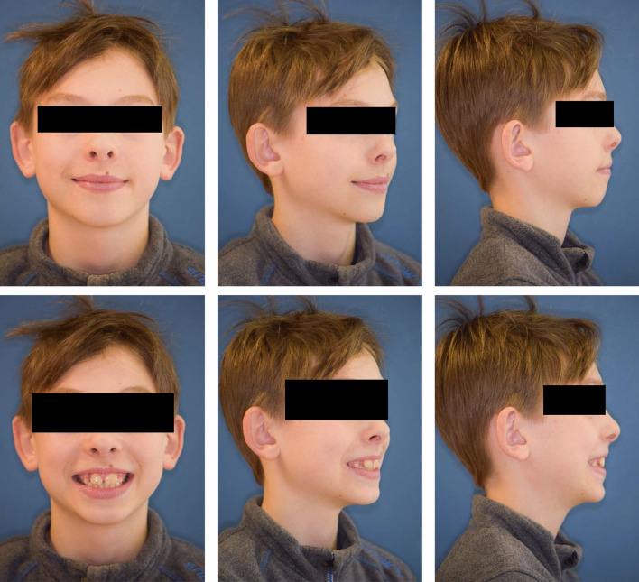
Tooth gemination is a dental phenomenon in which a single tooth bud attempts to divide into two, resulting in the formation of a structure that appears as two teeth but originates from the same follicle. This partial separation is often indicated clinically by a groove or depression that suggests the presence of two distinct teeth (Rajeswari M, Ananthalakshmi R. 2011. Gemination-case report and review. Indian Journal of Multidisciplinary Dentistry). The distinction between gemination and fusion plays an important role in treatment planning. If the number of teeth is one less, the tooth is fused and not geminated. In addition, it is assumed in the literature that geminated teeth have a single root canal and fused teeth have two separate root canals (Mahendra et al. in Case Rep Dent. 2014:425343, 2014;Duncan and Helpin in Oral Surg Oral Med Oral Pathol 64:82-87, 1987). The gemination of teeth is relatively rare and occurs mainly in the frontal region of the upper jaw. The prevalence of unilateral tooth gemination in the primary dentition is between 0.01 and 0.04% and in the permanent dentition: 0.05% (Duncan and Helpin in Oral Surg Oral Med Oral Pathol 64:82-87, 1987). Gemination management often requires a multidisciplinary approach and involves several steps ( Rajeswari M, Ananthalakshmi R. 2011. Gemination-case report and review. Indian Journal of Multidisciplinary Dentistry). The orthodontist will then take a thorough medical, dental and family history and perform clinical and radiographic examinations to confirm the diagnosis. Treatment options would include reshaping and restoring teeth with appropriate materials, performing root canal treatment followed by reduction of the mesiodistal width and crown restoration, extraction if the tooth is not suitable for root canal treatment followed by orthodontic space closure or fixed or removable prosthesis if required, transplantation of supernumerary teeth to replace the missing tooth. This case report presents a patient with gemination of teeth 11, 21 and progressive Class III growth tendency. In this case, the malformed anterior teeth were extracted and the gap was closed using skeletal anchorage. Patients with missing central incisors often require a complex interdisciplinary treatment, whether a prosthetic tooth-supported restoration of the missing anterior tooth, single implant, or orthodontic space closure are chosen. Ideally, each alternative should fulfill individual aesthetic concerns, functional requirements, and periodontal tissue health, not only at the end of treatment but also in the long term (Marco in Sem Orthodont 26:1, 2020; Rosa M, Zachrisson BU. Integrating space closure and esthetic dentistry in patients with missing maxillary lateral incisors. J Clin Orthod. 2007; 41(9); Czochrowska ,E.M.,Skaare,A.B.,Stevnik A, Zachrisson, B.U. Outcome of orthodontic space closure with a missing maxillary central incisor;) If gap closure is chosen, it is important to select the correct orthodontic appliance and anchorage especially in Class III patients with sagittal maxillary deficiency.
 求助内容:
求助内容: 应助结果提醒方式:


